

Vascular Floor of Mouth Mass with Unanticipated Intracranial, Orbital, and Vertebral Associated Involvements
- PMID: 30758759
- PMCID: PMC7021860
- DOI: 10.1007/s12105-019-01017-1
Vascular Floor of Mouth Mass with Unanticipated Intracranial, Orbital, and Vertebral Associated Involvements
Abstract
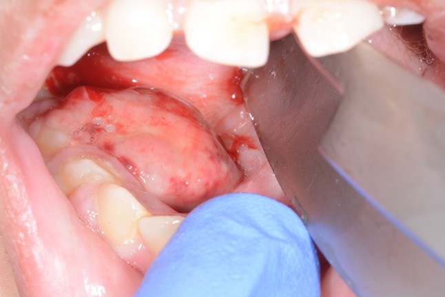
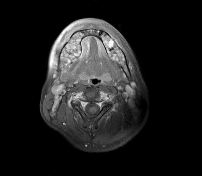
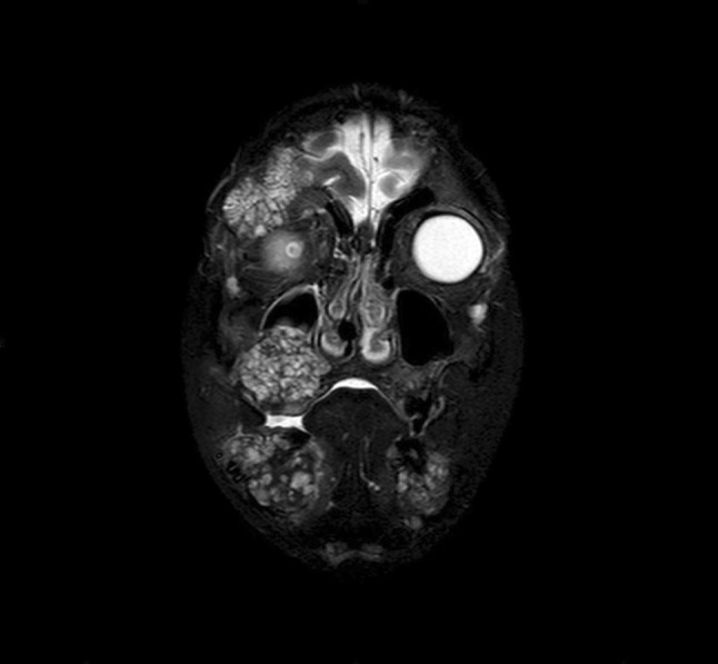
We report on a case in which a blanching, unobtrusive oral growth proved to be a systemic threat. A blind, epileptic child presented with a bleeding oral floor mass of 4 weeks. Biopsy showed small, dilated vascular spaces with reactive fibroblasts. MRI indicated distribution of expansile lesions in the mandible, cranial base, and right orbit that had possibly contributed to the patient's years-long neurologic deficits. A subsequent bone scan indicated lesions in multiple axial bones. Histologic markers confirmed the presentation of a rare cystic vascular pathology. Cystic Angiomatosis is a disease of intraosseous vascular malformations with occasional visceral involvements. Oral and craniomaxillofacial cases are especially rare and presentations can involve neuropsychiatric deficits, sensory issues, and mucosal bleeding. While clinicians are often dismissive of intraoral bleeding because of the prevalence of periodontal disease, careful evaluation is nonetheless critical to rule out underlying diseases with a possibly significant systemic involvement.
Keywords: Blindness; Cystic angiomatosis; Epilepsy; Vascular neoplasm.
Conflict of interest statement
The authors declare that they have no conflict of interest.
Figures
Similar articles
-
Skeletal angiomatosis - rare cause of bone destruction: a case report with review of literature.Indian J Pathol Microbiol. 2008 Oct-Dec;51(4):515-8. doi: 10.4103/0377-4929.43745. Indian J Pathol Microbiol. 2008. PMID: 19008580 Review.
-
Hemangiomatosis associated with osteolysis of the mandible in a dog resembling Gorham-Stout disease in humans.Vet Pathol. 2005 Jul;42(4):489-91. doi: 10.1354/vp.42-4-489. Vet Pathol. 2005. PMID: 16006608
-
Plunging arteriovenous malformation in the floor of the mouth: a case report.Br J Oral Maxillofac Surg. 2010 Dec;48(8):e35-7. doi: 10.1016/j.bjoms.2010.07.015. Epub 2010 Aug 21. Br J Oral Maxillofac Surg. 2010. PMID: 20728968
-
Oral intra vascular papillary endothelial hyperplasia in the floor of the mouth.Indian J Dent Res. 2004 Oct-Dec;15(4):149-51. Indian J Dent Res. 2004. PMID: 16035645
-
Massive craniofacial intraosseous vascular malformation resembling cystic angiomatosis: report of 2 cases and review of the literature.J Oral Maxillofac Surg. 2011 Jan;69(1):204-14. doi: 10.1016/j.joms.2010.07.072. Epub 2010 Nov 2. J Oral Maxillofac Surg. 2011. PMID: 21050648 Review.
References
-
- Hossein Mortazavi S, Khodayari A, Khojasteh A, Abbas FM, Mehrdad L, Kiani MT, et al. Massive craniofacial intraosseous vascular malformation resembling cystic angiomatosis: report of 2 cases and review of the literature. J Oral Maxillofac Surg. 2011;69:204–214. doi: 10.1016/j.joms.2010.07.072. - DOI - PubMed
-
- Beeram RK, Seelam S. Massive craniofacial cystic angiomatosis and extra-skeletal angiomatosis: a unique case report and brief review of literature. J Oral Maxillofac Surg, Med, Pathol. 2016;28:122–127. doi: 10.1016/j.ajoms.2015.08.008. - DOI
Publication types
MeSH terms
LinkOut - more resources
Full Text Sources
