

Morphometric analysis of spinal cord termination in Cavalier King Charles Spaniels
- PMID: 30758868
- PMCID: PMC6430917
- DOI: 10.1111/jvim.15437
Morphometric analysis of spinal cord termination in Cavalier King Charles Spaniels
Erratum in
-
Erratum for Morphometric analysis of spinal cord termination in Cavalier King Charles Spaniels.J Vet Intern Med. 2019 Jul;33(4):1840. doi: 10.1111/jvim.15514. Epub 2019 May 10. J Vet Intern Med. 2019. PMID: 31321833 Free PMC article. No abstract available.
Abstract
Background: There is an association between Chiari malformations, syringomyelia (CMSM) and tethered cord syndrome (TCS) in people, suggesting Cavalier King Charles Spaniels (CKCS) with CMSM could also have TCS. Currently there are no data on the position of the caudal spinal cord structures in CKCS.
Objective: To describe and compare location of spinal cord termination in CKCS with weight-matched controls and to examine the relationship between SM and spinal cord termination.
Animals: Thirty-nine CKCS and 33 controls with thoracolumbar MRIs; 34 of 39 CKCS also had cervical MRIs.
Methods: Blinded retrospective study. Spinal cord and dural sac termination were determined from T2-weighted sagittal and transverse images and half-Fourier acquisition single-shot turbo spin echo sequences. Intra-observer reliability was determined using kappa analysis. Presence of SM was compared with location of spinal cord and dural sac termination.
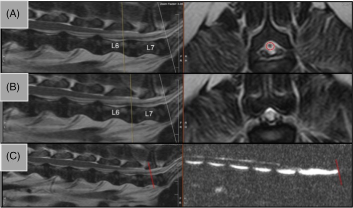
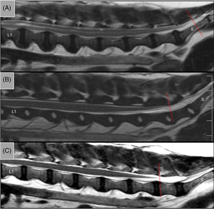
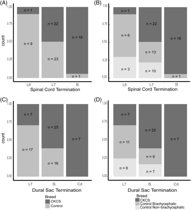
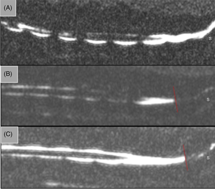
Results: Intra-observer reliability was moderate for identifying spinal cord termination (Kappa = 0.6) and good for dural sac termination (Kappa = 0.8). The spinal cord terminated at lumbar vertebra 6 (L6) in 1, 7 (L7) in 22, and sacrum in 16 CKCS versus 9 at L6, 23 at L7, 1 at sacrum in controls. Spinal cord (P < .001) and dural sac (P = .002) termination were significantly more caudal in CKCS compared to controls. The presence of thoracolumbar SM was associated with more caudal dural sac termination in CKCS (P = .03).
Conclusions and clinical importance: The relationship between TL SM and possible spinal cord tethering because of a more caudal dural sac termination should be investigated.
Keywords: conus medullaris; dural sac; filum terminale; syringomyelia.
© 2019 The Authors. Journal of Veterinary Internal Medicine published by Wiley Periodicals, Inc. on behalf of the American College of Veterinary Internal Medicine.
Conflict of interest statement
Authors declare no conflict of interest.
Figures
References
-
- Hechler AC, Moore SA. Understanding and treating Chiari‐like malformation and syringomyelia in dogs. Top Companion Anim Med. 2018;33:1‐11. - PubMed
-
- Parker JE, Knowler SP, Rusbridge C, Noorman E, Jeffery ND. Prevalence of asymptomatic syringomyelia in Cavalier King Charles Spaniels. Vet Rec. 2011;168:667‐667. - PubMed
-
- Cerda‐Gonzalez S, Olby NJ, Broadstone R, McCullough S, Osborne JA. Characteristics of cerebrospinal fluid flow in Cavalier King Charles Spaniels analyzed using phase velocity cine magnetic resonance imaging. Vet Radiol Ultrasound. 2009;50:467‐476. - PubMed
-
- Driver CJ, Volk HA, Rusbridge C, Van Ham LM. An Update on the pathogenesis of syringomyelia secondary to Chiari‐like malformations in dogs. Vet J. 2013;198:551‐559. - PubMed
-
- Fenn J, Schmidt MJ, Simpson H, Driver CJ, Volk HA. Venous sinus volume in the caudal cranial fossa in Cavalier King Charles Spaniels with syringomyelia. Vet J. 2013;197:896‐897. - PubMed
MeSH terms
Grants and funding
LinkOut - more resources
Full Text Sources
Medical