. 2019 Apr 1;316(4):L691-L700.
doi: 10.1152/ajplung.00282.2018. Epub 2019 Feb 13.

Extracellular vesicles: another compartment for the second messenger, cyclic adenosine monophosphate

Affiliations

Extracellular vesicles: another compartment for the second messenger, cyclic adenosine monophosphate

Sarah L Sayner et al. Am J Physiol Lung Cell Mol Physiol. .

Abstract

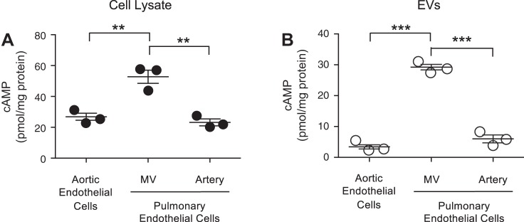
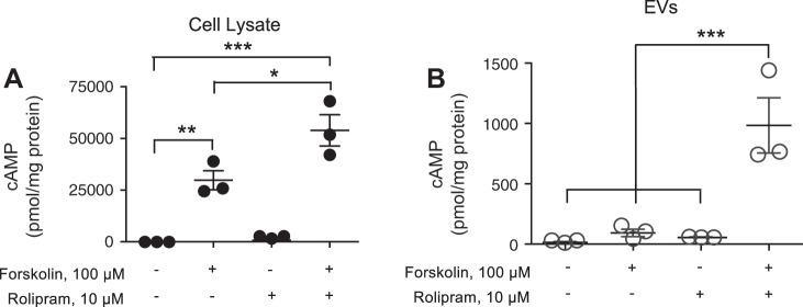
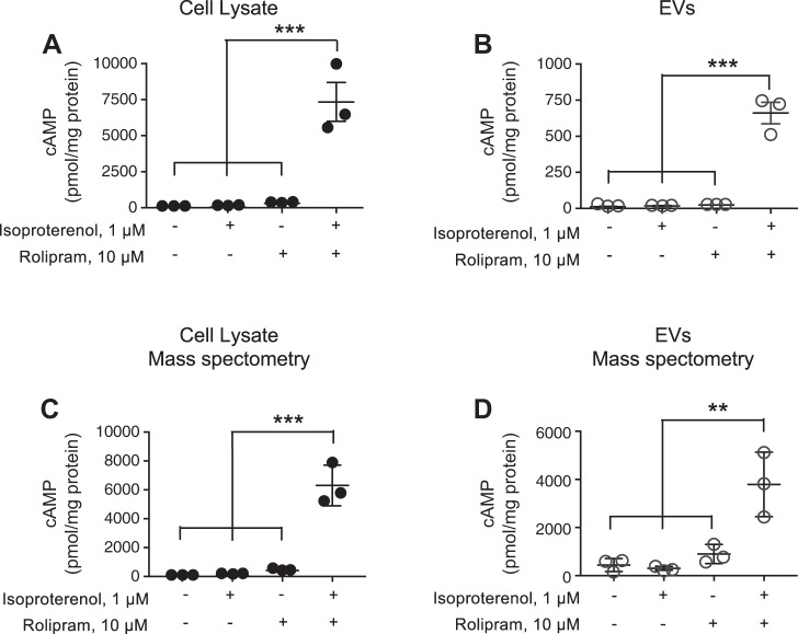
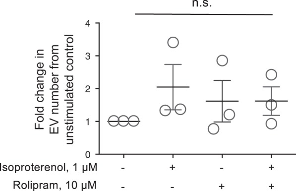
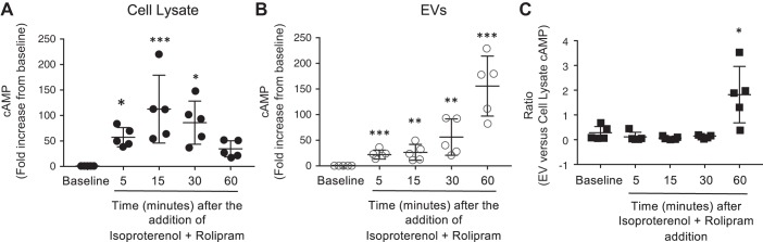
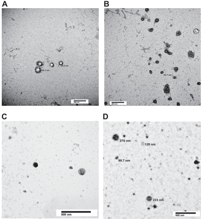
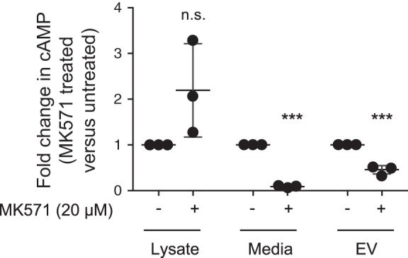
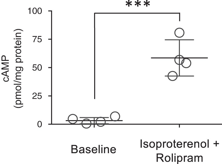
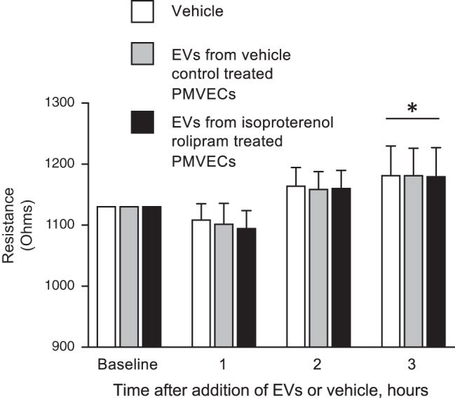
The second messenger, cAMP, is highly compartmentalized to facilitate signaling specificity. Extracellular vesicles (EVs) are submicron, intact vesicles released from many cell types that can act as biomarkers or be involved in cell-to-cell communication. Although it is well recognized that EVs encapsulate functional proteins and RNAs/miRNAs, currently it is unclear whether cyclic nucleotides are encapsulated within EVs to provide an additional second messenger compartment. Using ultracentrifugation, EVs were isolated from the culture medium of unstimulated systemic and pulmonary endothelial cells. EVs were also isolated from pulmonary microvascular endothelial cells (PMVECs) following stimulation of transmembrane adenylyl cyclase (AC) in the presence or absence of the phosphodiesterase 4 inhibitor rolipram over time. Whereas cAMP was detected in EVs isolated from endothelial cells derived from different vascular beds, it was highest in EVs isolated from PMVECs. Treatment of PMVECs with agents that increase near-membrane cAMP led to an increase in cAMP within corresponding EVs, yet there was no increase in EV number. Elevated cell cAMP, measured by whole cell measurements, peaked 15 min after treatment, yet in EVs the peak increase in cAMP was delayed until 60 min after cell stimulation. Cyclic AMP was also increased in EVs collected from the perfusate of isolated rat lungs stimulated with isoproterenol and rolipram, thus corroborating cell culture findings. When added to unperturbed confluent PMVECs, EVs containing elevated cAMP were not barrier disruptive like cytosolic cAMP but maintained monolayer resistance. In conclusion, PMVECs release EVs containing cAMP, providing an additional compartment to cAMP signaling.

Keywords: cyclic adenosine monophosphate; endothelium; extracellular vesicles; isolated lung; phosphodiesterase.


Conflict of interest statement

No conflicts of interest, financial or otherwise, are declared by the authors.

Figures

Fig. 1.
Fig. 1.
Extracellular vesicles (EVs) derived from endothelial cells of different vascular beds contain cAMP. cAMP was determined in whole cell lysate (A) and EVs (B) collected from unstimulated endothelial cells isolated from either the systemic circulation (aortic endothelial cells) or different vascular segments of pulmonary circulation, i.e., pulmonary endothelial cells: pulmonary microvascular endothelial cells (MV) or pulmonary artery endothelial cells (artery). EVs were isolated from the culture media, as described in methods. cAMP was normalized to protein content (n = 3; **P < 0.01 and ***P < 0.001, 1-way ANOVA with the Tukey post hoc test). ● and ○, Individual data points.
Fig. 2.
Fig. 2.
Direct activation of transmembrane adenylyl cyclase (AC) in PMVECs increases cellular and extracellular vesicle (EV)-cAMP. Pulmonary microvascular endothelial cells were stimulated with or without forskolin (100 µM) in the presence or absence of rolipram (10 µM) for 15 min and cell lysates and EV isolated. cAMP in the whole cell lysates (A) or EVs (B) was analyzed and normalized to protein content (n = 3; ***P < 0.001, **P < 0.01, and *P < 0.05, 1-way ANOVA with the Tukey post hoc test). ● and ○, Individual data points.
Fig. 3.
Fig. 3.
Isoproterenol activation of transmembrane adenylyl cyclase (AC) in pulmonary microvascular endothelial cells (PMVECs) increases cellular and extracellular vesicle (EV)-cAMP. PMVECs were stimulated for 15 min with or without isoproterenol in the presence or absence of rolipram and cell lysates (A and C) and EVs (B and D) analyzed for cAMP by either EIA (A and B) or mass spectrometry (C and D). cAMP was normalized to protein content (n = 3; ***P < 0.001, **P < 0.01, 1-way ANOVA with the Tukey post hoc test). ● and ○, Individual data points.
Fig. 4.
Fig. 4.
Isoproterenol activation of transmembrane adenylyl cyclase (AC) does not increase the number of extracellular vesicle (EV) released. Pulmonary microvascular endothelial cells were stimulated without or with isoproterenol (1 µM) in the presence or absence of rolipram (10 µM), and EVs were isolated as described in materials and methods. EV counts were determined by flow cytometry of particles <1 µM in diameter [n = 3; not significant (NS), 1-way ANOVA with the Tukey post hoc test]. ○, Individual data points.
Fig. 5.
Fig. 5.
Agonist stimulation increases extracellular vesicle (EV)-cAMP over time. A and B: pulmonary microvascular endothelial cells were stimulated with isoproterenol and rolipram over time (0–60 min), and cell lysates (A) and EVs (B) were analyzed for cAMP and normalized to protein concentration. C: the ratio of cAMP/mg protein from EVs vs. cell lysate for each time point (n = 5; ***P < 0.001, **P < 0.01, and *P < 0.05 vs. baseline, Student’s t-test). ●, ○, and ■, Individual data points.
Fig. 6.
Fig. 6.
Transmission electron microscopy (TEM) analysis of extracellular vesicles (EVs). EVs isolated from either unstimulated or isoproterenol- (1 µM) and rolipram-stimulated (10 µM) (60 min) pulmonary microvascular endothelial cells were fixed and counterstained for TEM. EVs from both unstimulated (A and B) and isoproterenol- and rolipram-stimulated (C and D) pulmonary microvascular endothelial cells exhibit characteristics of intact, heterogenous vesicles. The EVs range in size from exosomes (≤150 nm) to microparticles (≥200 nm up to 1 μM). Representative images from 6 independent preparations for each condition.
Fig. 7.
Fig. 7.
MK571 attenuates extracellular vesicle (EV) cAMP as well as cAMP efflux. Pulmonary microvascular endothelial cells were pretreated with or without MK571 (20 µM) for 5 min before stimulation with isoproterenol (1 µM) and rolipram (10 µM) for 60 min. For pretreated cells, MK571 (20 µM) was maintained throughout the isoproterenol and rolipram treatment. The cell lysate, media, and EVs were collected for cAMP and protein analysis and the ratio of untreated vs. MK571 treated levels of cAMP normalized to protein calculated for each experimental condition [n = 3; ***P < 0.01 vs. without MK571 and not significant (NS); Student’s t-test]. ●, Individual data points.
Fig. 8.
Fig. 8.
Extracellular vesicles (EVs) from isolated rat lungs perfused with isoproterenol and rolipram contain elevated cAMP. Isolated rat lungs were perfused with or without (i.e vehicle control) isoproterenol (1 µM) and rolipram (10 µM) for 1 h and the perfusate collected for isolation of EVs as described in materials and methods. EVs were analyzed for cAMP and normalized to protein concentration (n = 4 separate lungs/experimental condition; *P < 0.001 vs. vehicle control, Student’s t-test). ○, Individual data points.
Fig. 9.
Fig. 9.
Extracellular vesicles (EVs) from unstimulated (vehicle control) as well as isoproterenol and rolipram stimulated pulmonary microvascular endothelial cells (PMVECs) do not alter transendothelial electrical resistance. EVs collected (as described in materials and methods) from PMVECs stimulated with isoproterenol (1 µM) and rolipram (10 µM) or vehicle control for 60 min were applied to confluent PMVEC monolayers and the change in resistance recorded over time. DMEM was used as the EV vehicle control. EVs collected from vehicle control or isoproterenol- and rolipram-treated PMVECs did not alter monolayer resistance from vehicle control-treated PMVECs (data represent n = 4, with each individual experiment performed as n = 2 or 3; *P < 0.05, 2-way ANOVA with Tukey post hoc test).

References

    1. Abid Hussein MN, Böing AN, Sturk A, Hau CM, Nieuwland R. Inhibition of microparticle release triggers endothelial cell apoptosis and detachment. Thromb Haemost 98: 1096–1107, 2007. doi:10.1160/TH05-04-0231. - DOI - PubMed
    1. Amabile N, Guignabert C, Montani D, Yeghiazarians Y, Boulanger CM, Humbert M. Cellular microparticles in the pathogenesis of pulmonary hypertension. Eur Respir J 42: 272–279, 2013. doi:10.1183/09031936.00087212. - DOI - PubMed
    1. Bähre H, Hartwig C, Munder A, Wolter S, Stelzer T, Schirmer B, Beckert U, Frank DW, Tümmler B, Kaever V, Seifert R. cCMP and cUMP occur in vivo. Biochem Biophys Res Commun 460: 909–914, 2015. doi:10.1016/j.bbrc.2015.03.115. - DOI - PMC - PubMed
    1. Bakouboula B, Morel O, Faure A, Zobairi F, Jesel L, Trinh A, Zupan M, Canuet M, Grunebaum L, Brunette A, Desprez D, Chabot F, Weitzenblum E, Freyssinet JM, Chaouat A, Toti F. Procoagulant membrane microparticles correlate with the severity of pulmonary arterial hypertension. Am J Respir Crit Care Med 177: 536–543, 2008. doi:10.1164/rccm.200706-840OC. - DOI - PubMed
    1. Bastarache JA, Fremont RD, Kropski JA, Bossert FR, Ware LB. Procoagulant alveolar microparticles in the lungs of patients with acute respiratory distress syndrome. Am J Physiol Lung Cell Mol Physiol 297: L1035–L1041, 2009. doi:10.1152/ajplung.00214.2009. - DOI - PMC - PubMed

Publication types

LinkOut - more resources