Review
. 2019 May 1;316(5):C583-C604.
doi: 10.1152/ajpcell.00523.2018. Epub 2019 Feb 13.

Posttranslational modifications of mitochondrial fission and fusion proteins in cardiac physiology and pathophysiology

Affiliations
Review

Posttranslational modifications of mitochondrial fission and fusion proteins in cardiac physiology and pathophysiology

Stephanie M Adaniya et al. Am J Physiol Cell Physiol. .

Abstract

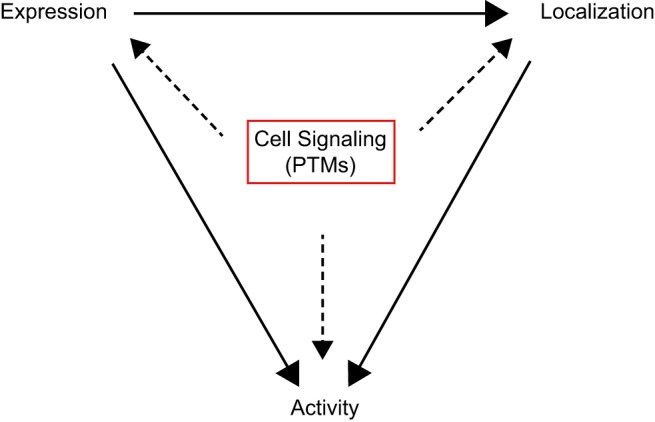
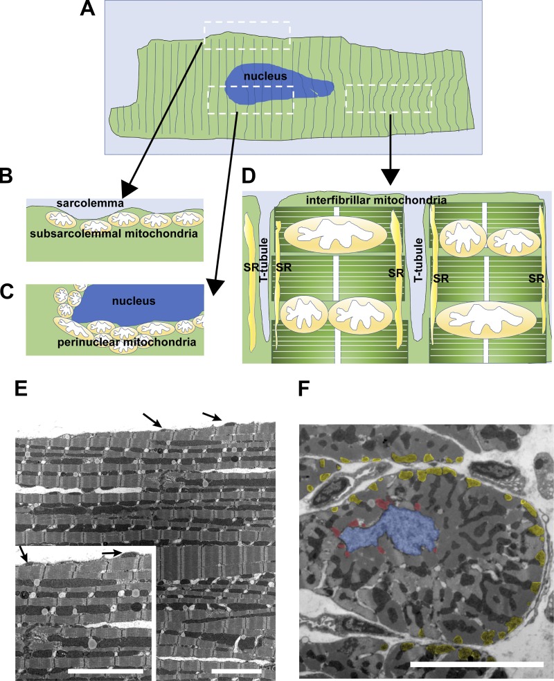
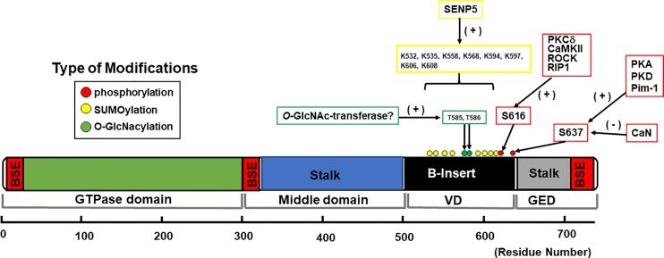
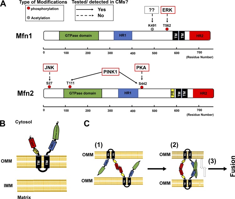
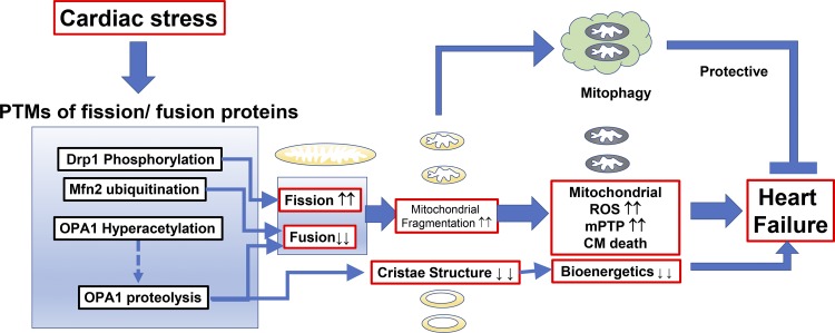
Mitochondrial fragmentation frequently occurs in chronic pathological conditions as seen in various human diseases. In fact, abnormal mitochondrial morphology and mitochondrial dysfunction are hallmarks of heart failure (HF) in both human patients and HF animal models. A link between mitochondrial fragmentation and cardiac pathologies has been widely proposed, but the physiological relevance of mitochondrial fission and fusion in the heart is still unclear. Recent studies have increasingly shown that posttranslational modifications (PTMs) of fission and fusion proteins are capable of directly modulating the stability, localization, and/or activity of these proteins. These PTMs include phosphorylation, acetylation, ubiquitination, conjugation of small ubiquitin-like modifier proteins, O-linked-N-acetyl-glucosamine glycosylation, and proteolysis. Thus, understanding the PTMs of fission and fusion proteins may allow us to understand the complexities that determine the balance of mitochondrial fission and fusion as well as mitochondrial function in various cell types and organs including cardiomyocytes and the heart. In this review, we summarize present knowledge regarding the function and regulation of mitochondrial fission and fusion in cardiomyocytes, specifically focusing on the PTMs of each mitochondrial fission/fusion protein. We also discuss the molecular mechanisms underlying abnormal mitochondrial morphology in HF and their contributions to the development of cardiac diseases, highlighting the crucial roles of PTMs of mitochondrial fission and fusion proteins. Finally, we discuss the future potential of manipulating PTMs of fission and fusion proteins as a therapeutic strategy for preventing and/or treating HF.

Keywords: DLP1; Drp1; Mfn; OPA1; mitophagy.


Conflict of interest statement

No conflicts of interest, financial or otherwise, are declared by the authors.

Figures

Fig. 1.
Fig. 1.
Regulation of mitochondrial morphology by posttranslational modifications (PTMs) of mitochondrial fission and fusion proteins. Schematic model of the three main regulators (protein expression, localization at the mitochondria, and overall GTPase activity) of mitochondrial fission and fusion, which interdependently modulate the balance of fission and fusion.
Fig. 2.
Fig. 2.
Subpopulations of mitochondria in adult cardiomyocytes. A: illustration of a single adult cardiomyocyte and diagram of different subpopulations of mitochondria. B: illustration of longitudinal section of the area indicated in A including subsarcolemmal mitochondria. C: illustration of longitudinal section of the area indicated in A including perinuclear mitochondria. D: illustration of longitudinal section of the area indicated in A including interfibrillar mitochondria. Note that interfibrillar mitochondria face the sarcoplasmic reticulum (SR). T tubule, transverse tubule. E: transmission electron microscope image of the longitudinal section of a papillary muscle obtained from an adult rabbit ventricle. Arrows show the location of subsarcolemmal mitochondria. Scale bars, 5 µm. F: transmission electron microscope image of the short axis of a papillary muscle obtained from an adult rabbit ventricle. Blue area indicates location of the nucleus. Red areas indicate perinuclear mitochondria. Yellow areas indicate subsarcolemmal mitochondria. Scale bar, 5 µm.
Fig. 3.
Fig. 3.
Schematic diagram of dynamin-related protein-1 (Drp1) structure and distribution of posttranslational modifications in cardiomyocytes and hearts. Structure-based domain architecture of human Drp1 isoform 1 is shown on the basis of the published data of Drp1 isoform 2 given by Fröhlich et al. (64). All the posttranslational modifications listed in this diagram are based on reports using native cardiomyocytes or heart tissues (see also Table 3). Information on sites of conjugation of small ubiquitin-like modifier (SUMO) proteins (SUMOylation) was collected from PhosphoSitePlus (80). BSE, bundle signaling element; CaN, Ca2+-dependent phosphatase calcineurin; GED, GTPase effector domain; O-GlcNAc, O-linked-N-acetyl-glucosamine; O-GlycNAcylation, O-linked-N-acetyl-glucosamine glycosylation; Pim-1, proto-oncogene serine/threonine-protein kinase; RIP1, receptor-interacting protein-1; ROCK, Rho-associated protein kinase; SENP5, sentrin/SUMO-specific protease 5; VD, variable domain.
Fig. 4.
Fig. 4.
Schematic diagram of structure and distribution of posttranslational modifications of mitofusin 1 and 2 (Mfn1 and Mfn2, respectively), and mechanism of outer mitochondrial membrane (OMM) fusion by Mfn proteins. A: structure-based domain architectures of human Mfn1 and Mfn2 are depicted on the basis of the published data given by Franco et al. (; see also Table 4). TM, transmembrane domain; HR, heptad-repeat domain. B: Mfn2 topology at the OMM, with two TM domains, a small bridge structure at the intermembrane space, and large structures at the cytosolic space. C: representative cartoon shows Mfn2-Mfn2 interaction. Initial tethering is mediated between heptad-repeat 2 domains (HR2s) from the opposite OMM (step 1). Next, dimerization of GTPase domains occurs (step 2), and power stroke from GTPase hydrolysis promotes OMM fusion (step 3). CMs, cardiomyocytes; IMM, inner mitochondrial membrane; PINK1, phosphatase and tensin homolog (PTEN)-induced putative kinase 1; PR, proline-rich domain.
Fig. 5.
Fig. 5.
Schematic diagram of optic atrophy protein-1 (OPA1) structure and variant-dependent proteolysis. A: structure-based domain architecture of human OPA1 is depicted. Numbers in the domains indicate the exon number. Red exons are involved in the formation of splice variants. B: variant-dependent proteolysis and the structures of the membrane-anchored long and soluble short forms of OPA1 (L-OPA1 and S-OPA1, respectively). C: summary table of variant-dependent proteolysis. GED, GTPase effector domain; IMM, inner mitochondrial membrane; IMS, intermembrane space; MPP, mitochondrial processing peptidase; MTS, mitochondrial-targeting sequence; OMA1, overlapping with the m-AAA protease 1 homolog; S1 and S2, sites 1 and 2; TM, transmembrane domain; YME1L, i-AAA metalloprotease.
Fig. 6.
Fig. 6.
Posttranslational modifications (PTMs) of mitochondrial fission and fusion proteins by cardiac pathological signaling promote heart failure but also serve as compensatory mechanisms for protecting cardiomyocytes (CMs). DLP1, dynamin-related protein-1; Mfn2, mitofusin 2; mPTP, mitochondrial permeability transition pore; OPA1, optic atrophy protein-1; ROS, reactive oxygen species.

References

    1. Acin-Perez R, Lechuga-Vieco AV, Del Mar Muñoz M, Nieto-Arellano R, Torroja C, Sánchez-Cabo F, Jiménez C, González-Guerra A, Carrascoso I, Benincá C, Quiros PM, López-Otín C, Castellano JM, Ruíz-Cabello J, Jiménez-Borreguero LJ, Enríquez JA. Ablation of the stress protease OMA1 protects against heart failure in mice. Sci Transl Med 10: eaan4935, 2018. doi:10.1126/scitranslmed.aan4935. - DOI - PubMed
    1. Alexander C, Votruba M, Pesch UE, Thiselton DL, Mayer S, Moore A, Rodriguez M, Kellner U, Leo-Kottler B, Auburger G, Bhattacharya SS, Wissinger B. OPA1, encoding a dynamin-related GTPase, is mutated in autosomal dominant optic atrophy linked to chromosome 3q28. Nat Genet 26: 211–215, 2000. doi:10.1038/79944. - DOI - PubMed
    1. Anand R, Wai T, Baker MJ, Kladt N, Schauss AC, Rugarli E, Langer T. The i-AAA protease YME1L and OMA1 cleave OPA1 to balance mitochondrial fusion and fission. J Cell Biol 204: 919–929, 2014. doi:10.1083/jcb.201308006. - DOI - PMC - PubMed
    1. Aon MA, Cortassa S, O’Rourke B. Redox-optimized ROS balance: a unifying hypothesis. Biochim Biophys Acta 1797: 865–877, 2010. doi:10.1016/j.bbabio.2010.02.016. - DOI - PMC - PubMed
    1. Archer SL. Mitochondrial fission and fusion in human diseases. N Engl J Med 370: 1074, 2014. doi:10.1056/NEJMc1316254. - DOI - PubMed

Publication types

MeSH terms

Substances