. 2019 Feb 13;14(2):e0211151.
doi: 10.1371/journal.pone.0211151. eCollection 2019.

The primate-specific peptide Y-P30 regulates morphological maturation of neocortical dendritic spines

Affiliations

The primate-specific peptide Y-P30 regulates morphological maturation of neocortical dendritic spines

Janine R Neumann et al. PLoS One. .

Abstract

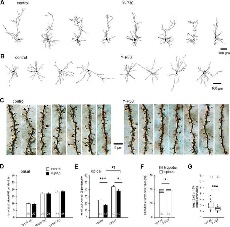
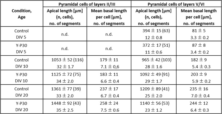
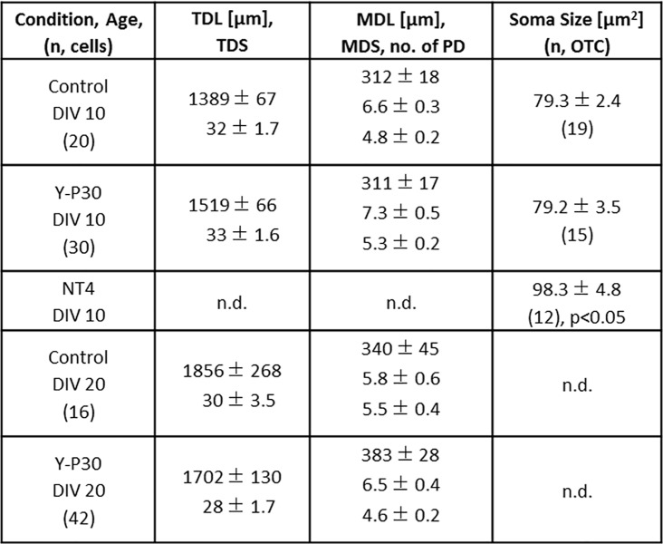
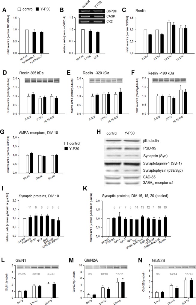
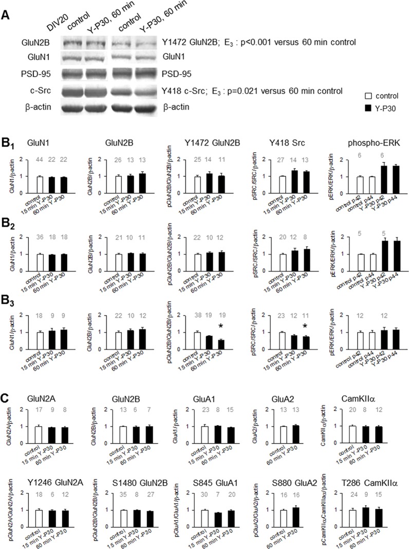
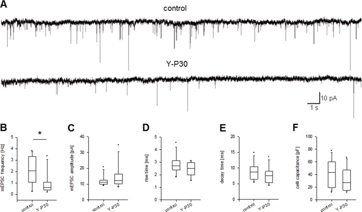
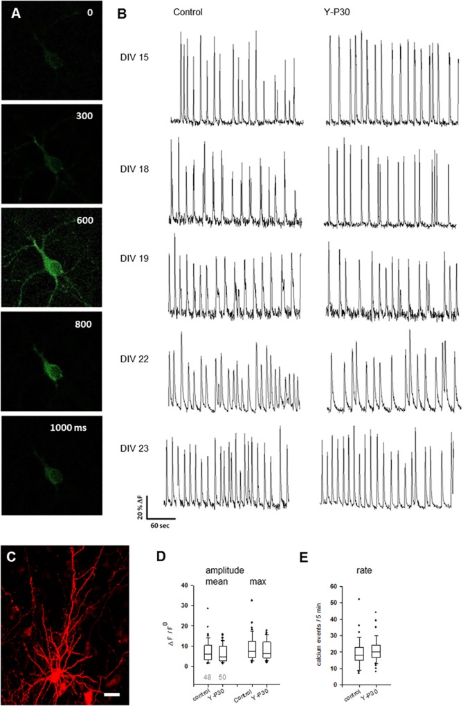
The 30-amino acid peptide Y-P30 corresponds to the N-terminus of the primate-specific, sweat gland-derived dermcidin prepropeptide. Previous work has revealed that Y-P30 enhances the interaction of pleiotrophin and syndecans-2/3, and thus represents a natural ligand to study this signaling pathway. In immature neurons, Y-P30 activates the c-Src and p42/44 ERK kinase pathway, increases the amount of F-actin in axonal growth cones, and promotes neuronal survival, cell migration and axonal elongation. The action of Y-P30 on axonal growth requires syndecan-3 and heparan sulfate side chains. Whether Y-P30 has the potential to influence dendrites and dendritic protrusions has not been explored. The latter is suggested by the observations that syndecan-2 expression increases during postnatal development, that syndecan-2 becomes enriched in dendritic spines, and that overexpression of syndecan-2 in immature neurons results in a premature morphological maturation of dendritic spines. Here, analysing rat cortical pyramidal and non-pyramidal neurons in organotypic cultures, we show that Y-P30 does not alter the development of the dendritic arborization patterns. However, Y-P30 treatment decreases the density of apical, but not basal dendritic protrusions at the expense of the filopodia. Analysis of spine morphology revealed an unchanged mushroom/stubby-to-thin spine ratio and a shortening of the longest decile of dendritic protrusions. Whole-cell recordings from cortical principal neurons in dissociated cultures grown in the presence of Y-P30 demonstrated a decrease in the frequency of glutamatergic mEPSCs. Despite these differences in protrusion morphology and synaptic transmission, the latter likely attributable to presynaptic effects, calcium event rate and amplitude recorded in pyramidal neurons in organotypic cultures were not altered by Y-P30 treatment. Together, our data suggest that Y-P30 has the capacity to decelerate spinogenesis and to promote morphological, but not synaptic, maturation of dendritic protrusions.


Conflict of interest statement

The authors have declared that no competing interests exist.

Figures

Fig 1
Fig 1. Y-P30 does not alter somatodendritic differentiation of cortical neurons but promotes filopodia pruning and morphological maturation of spines on pyramidal neurons.
(A) Chronic treatment with Y-P30 had no effect on pyramidal cell dendritic growth; gallery of 4 representative cells shown per condition at DIV 10; for quantitative data see Fig 2, for graphs see S1 Fig. (B) Chronic treatment with Y-P30 had no effect on interneuronal dendritic growth; gallery of 4 representative cells shown per condition at DIV10; for quantitative data see Fig 3, for graphs see S2 Fig. (C) Segments of secondary oblique branches from the apical dendrite of seven 20 DIV pyramidal cells from control OTC and OTC treated with Y-P30. EGFP transfection at DIV 14; Y-P30 treatment with 1 μM daily from 15–19 DIV, controls were mock-stimulated daily with an equivalent volume of vehicle. Segments were ordered from left-to-right from high-to-low protrusion density to document variability; note the longer protrusions in the control group. (D) Y-P30 did not alter the density of interneuronal (IN) dendritic protrusions and basal dendritic protrusions of pyramidal cells (PC) at DIV 10 (treated with 1 μM Y-P30 at DIV 7 and DIV 9) and DIV 20 (daily one pulse of 1 μM Y-P30 from DIV 15–19). Plotted is the mean number ± SEM per 100 μm dendritic length. (E) Y-P30 decreased the density of protrusions on secondary oblique branches from the apical dendrites of pyramidal cells at DIV 10 by 27% and at DIV 20 by 13%. Plotted is the mean number ± SEM per 100 μm dendritic length. Note, that the protrusion density increased between DIV 10 and DIV 20 in vehicle-treated and in Y-P30 treated neurons. The number of cells analyzed is indicated in the bars; they derived from 9–12 OTC from three independent preparations; *** p<0.001, * p = 0.017, *1 p = 0.02, Mann-Whitney U-test. (F) Y-P30 increased the proportion of spines with heads (mushroom/stubby and thin spines) on secondary oblique branches from the apical dendrites of pyramidal cells at 20 DIV and decreased the proportion of filopodia (* p = 0.035, Mann-Whitney U-test). Numbers in the bars indicate the number of cells analyzed each with arbitrarily selected 3–4 segments each of about 50 μm length. (G) Assessment of protrusion length on secondary oblique branches from the apical dendrites of pyramidal cells at DIV 20. Plotted are the 10% longest protrusions sampled from 2–3 dendritic segments; every value is the mean of one cell, the numbers above the boxes are the number of cells analyzed. *** p<0.001, Mann-Whitney U-test.
Fig 2
Fig 2. Morphometric data of layer II/III and V/VI pyramidal neurons.
Cells reconstructed from DIV 5, DIV 10 and DIV 20 OTC; chronically exposed to Y-P30 or vehicle (control). Mean ± SEM, the number of cells (n) is given in brackets. At DIV 5, only cells from infragranular layers were analyzed because supragranular neurons are too immature to reliably identify the type. n.d., not determined.
Fig 3
Fig 3. Morphometric data of multipolar interneurons.
Cells reconstructed from DIV 10 and DIV 20 OTC; chronically exposed to Y-P30 or vehicle. Mean ± SEM for TDL, total dendritic length; TDS, total dendritic segments; MDL, mean dendritic length; MDS, mean dendritic segments; no.of PD, number of primary dendrites/neuron. The number of cells is given in brackets. Soma size (area in μm2 as proxy) of GABA-ir neurons is the grand average of somata analyzed in 19 and 15 OTC by sampling 50–80 somata in a perpendicular strip through all layers in every OTC. 20 ng/mL NT4 treatment has been done at DIV 10 as positive control (12 OTC) to show responsiveness of interneurons. ANOVA on ranks followed by Mann-Whitney U-test versus DIV 10 control p<0.05. n.d. not determined.
Fig 4
Fig 4. Y-P30 effects on expression of synaptic proteins.
(A) Y-P30 did not alter the mRNA expression of syndecan-2/-3 and of (B) CASK and casein kinase 2α (CK2) as determined with qPCR. Mean ± SEM of four (A) and three (B) independent experiments normalized to the expression of glucose-6-phosphate dehydrogenase (G6PDH) or 18S rRNA. (C) Y-P30 initially decreased the mRNA expression of reelin at DIV 3–5, as determined with RT-PCR, but no longer at DIV 10 and DIV 15. Mean ± SEM of six to ten qPCR experiments with cultures from four independent preparations normalized to the expression of G6PDH. For DIV 3, control level at DIV 3 was set to 1. Expression levels at DIV 5, 10 and 15 were expressed relative to the DIV 5 control level which was set to 1. (D-F) The amount of the full length reelin protein at ~385 kDa (D) and the proteolytic fragments at 320 kDa (E) and 180 kDa (F) were not altered after stimulation of cortical cultures with Y-P30. Mean ± SEM of up to ten lysates from four independent preparations normalized to the expression of βIII-tubulin; average control level at DIV 3 was set to 1. (G) The mRNA expression of GluA1, GluA2, and GluA3 determined with RT-PCR was unchanged in Y-P30-treated cortical OTC. Mean ± SEM of more than 12 reactions run with 4–5 cDNA libraries each synthesized with the mRNA isolated from 5 OTC; the average control levels were set to 1. (H-K) The expression of the pre- and postsynaptic proteins synapsin-1, S9 phosphorylated synapsin, synaptotagmin-1, synaptotagmin-2, synaptophysin, synaptopodin, PSD-95, glutamate decarboxylase GAD65/67, and GABAA receptor α1 subunit was unchanged in OTC treated with Y-P30 until DIV 10 (H, I) or DIV 15, DIV 18, DIV 20 (pooled). Graphs in I, K show mean ± SEM, the number of lysates (1 OTC/lysate) probed for every protein is given above the bars; note that an equal number of vehicle-stimulated control OTC was run. OTC from 3–5 independent culture preparations; normalization to the expression of βIII-tubulin or β-actin on the same filter membrane to correct for loading. Average control levels were set to 1, but only one control bar for all has been plotted and therefore, a SEM is not indicated in the white bars of I and K. (L-N) Chronic exposure of OTCs to Y-P30 did not alter the developmental expression levels of GluN1, GluN2A and GluN2B protein, as seen at DIV 5, DIV 10 and DIV 15. Mean ± SEM, number of lysates probed for every protein is given above the bars; OTC from five to six independent preparations; normalization to βIII-tubulin; average of the vehicle-stimulated control levels at DIV5 were set to 1.
Fig 5
Fig 5. Effects of Y-P30 on expression and phosphorylation of glutamate receptors, Src, and ERK.
(A-C) Qualitative (A) and quantitative (B, C) results of 15 and 60 min single-pulse treatment with Y-P30 at DIV 5, DIV 10 and DIV 20. (A) Representative blot at DIV 20; per lane, 50% of a lysate from a vehicle-treated control and a 60 min Y-P30-treated OTC was loaded on the left and the right side of a gel. Strips overlapping the target protein size range were cut after blotting from the nitrocellulose membrane and developed with antibodies to the indicated antigens. Note the reduction of Y1472 GluN2B and Y418 c-Src phosphorylation in the Y-P30-treated OTC (rightmost lane). (B1, B2, B3) Expression of GluN1, GluN2B, Y1472 phosphorylated GluN2B, Y418 phosphorylated c-Src, and phosphorylated p42/p44 ERK, at 15 min and 60 min after a single pulse of Y-P30. B1 top row at DIV 5; B2 middle row at DIV 10; B3 lower row at DIV 20. Note that p42/p44 ERK and c-Src phosphorylation were increased at DIV 5 and DIV 10. By contrast, at DIV 20, p42/p44 ERK was no longer activated, and Y418 c-Src and Y1472 GluN2B phosphorylation were decreased at 15 and at 60 min; Mann-Whitney U test p values given in (A). Mean ± SEM, number of lysates probed for every protein or phosphorylation site is given above the bars; vehicle-stimulated controls at the two time points have been pooled; OTC from five to six independent preparations. (C) Expression at DIV 20 of GluN2A and Y1246 GluN2A, GluN2B and S1480 GluN2B, GluA1 and S845 GluA1, GluA2 and S880 GluA2, and CamKIIα and T286 CamKIIα were not altered by a single pulse of Y-P30. Mean ± SEM, number of lysates probed for every protein or phosphorylation site is given above the bars; vehicle-stimulated controls at the two time points have been pooled; OTC from five to six independent preparations.
Fig 6
Fig 6. Y-P30 effects on neurotrophin expression.
(A) Y-P30 did not alter the expression of BDNF, NT4, NGF, TrkB and TrkC (determined for the kinase domain-bearing full length receptors), but quickly reduced NT3 mRNA. (B) The expression of NT3 mRNA was reduced at 1 and 2 h after a single pulse of Y-P30. (C) Longterm treatment with Y-P30, 3 pulses from DIV 5 to DIV 10 followed by analysis at least 12 h after the last pulse did not alter the levels of trophic factor and receptor mRNAs. Shown are means (in A) and mean ± SEM (bar graphs in B, C) normalized to G6PDH of six RT-PCR reactions performed with three cDNA libraries per time point each synthesized with mRNA isolated from 5 OTC; untreated control (0 min) was set to 1, * p<0.05 at 1 and 2h, two-tailed t-test.
Fig 7
Fig 7. Lower frequency of glutamatergic mEPSCs but no alteration in mEPSC amplitude and kinetics in cortical pyramidal neurons exposed to Y-P30.
(A) Sample traces of miniature excitatory postsynaptic currents (mEPSCs) recorded from DIV 15–17 dissociated cortical neurons under control conditions and following chronic exposure to Y-P30. (B) Recordings from neurons grown in the presence of Y-P30 revealed significantly lower mean mEPSC frequency, but not (C) mEPSC amplitude, (D) mEPSC rise time, or (E) mEPSC decay time. (F) Chronic exposure to Y-P30 did not have a significant effect on cell membrane capacitance. *p < 0.05, Mann-Whitney U-test.
Fig 8
Fig 8. Rate and amplitude of calcium events are not affected in pyramidal neurons in OTC exposed to Y-P30.
(A) GCaMP6m calcium signal of a DIV 14 pyramidal neuron. (B) Calcium event traces recorded at DIV 14–23 pyramidal neurons, vehicle-treated control and Y-P30 treated from DIV 8 with one pulse daily at 1 μM final concentration. (C) A representative cortical pyramidal cell at DIV 18 as identified by mCherry fluorescence, scale 20 μm. (D) Mean and maximal amplitude of calcium events were not different; Mann-Whitney U-test, p = 0.378 and p = 0.466 for mean and max amplitudes. (E) Rates of calcium events during a 5 min recording were not different, Mann-Whitney U-test, p = 0.126. Numbers in D indicate the number of total neurons recorded and plotted. Neurons were from 15 vehicle-treated and 14 Y-P30-treated OTC from 3 independent preparations.

Similar articles

Cited by

References

    1. Huttenlocher PR. Morphometric study of human cerebral cortex development. Neuropsychologia. 1990;28(6):517–27. - PubMed
    1. Petanjek Z, Judas M, Simic G, Rasin MR, Uylings HB, Rakic P, et al. Extraordinary neoteny of synaptic spines in the human prefrontal cortex. Proc Natl Acad Sci U S A. 2011; 108(32):13281–6. 10.1073/pnas.1105108108 - DOI - PMC - PubMed
    1. Penzes P, Cahill ME, Jones KA, VanLeeuwen JE, Woolfrey KM. Dendritic spine pathology in neuropsychiatric disorders. Nat Neurosci. 2011;14(3):285–93. 10.1038/nn.2741 - DOI - PMC - PubMed
    1. Schittek B, Hipfel R, Sauer B, Bauer J, Kalbacher H, Stevanovic S et al. Dermcidin: a novel human antibiotic peptide secreted by sweat glands. Nat Immunol. 2001;2(12):1133–37. 10.1038/ni732 - DOI - PubMed
    1. Schittek B The multiple facets of dermcidin in cell survival and host defense. J Innate Immun. 2012;4(4):349–60. 10.1159/000336844 - DOI - PMC - PubMed

Publication types