. 2019 Feb 13;14(2):e0210663.
doi: 10.1371/journal.pone.0210663. eCollection 2019.

Late effects of total body irradiation on hematopoietic recovery and immune function in rhesus macaques

Affiliations

Late effects of total body irradiation on hematopoietic recovery and immune function in rhesus macaques

Laura P Hale et al. PLoS One. .

Abstract

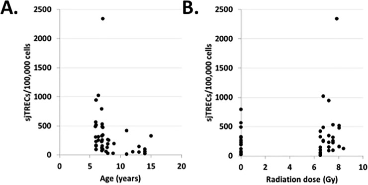
While exposure to radiation can be lifesaving in certain settings, it can also potentially result in long-lasting adverse effects, particularly to hematopoietic and immune cells. This study investigated hematopoietic recovery and immune function in rhesus macaques Cross-sectionally (at a single time point) 2 to 5 years after exposure to a single large dose (6.5 to 8.4 Gray) of total body radiation (TBI) derived from linear accelerator-derived photons (2 MeV, 80 cGy/minute) or Cobalt 60-derived gamma irradiation (60 cGy/min). Hematopoietic recovery was assessed through measurement of complete blood counts, lymphocyte subpopulation analysis, and thymus function assessment. Capacity to mount specific antibody responses against rabies, Streptococcus pneumoniae, and tetanus antigens was determined 2 years after TBI. Irradiated macaques showed increased white blood cells, decreased platelets, and decreased frequencies of peripheral blood T cells. Effects of prior radiation on production and export of new T cells by the thymus was dependent on age at the time of analysis, with evidence of interaction with radiation dose for CD8+ T cells. Irradiated and control animals mounted similar mean antibody responses to proteins from tetanus and rabies and to 10 of 11 serotype-specific pneumococcal polysaccharides. However, irradiated animals uniformly failed to make antibodies against polysaccharides from serotype 5 pneumococci, in contrast to the robust responses of non-irradiated controls. Trends toward decreased serum levels of anti-tetanus IgM and slower peak antibody responses to rabies were also observed. Taken together, these data show that dose-related changes in peripheral blood cells and immune responses to both novel and recall antigens can be detected 2 to 5 years after exposure to whole body radiation. Longer term follow-up data on this cohort and independent validation will be helpful to determine whether these changes persist or whether additional changes become evident with increasing time since radiation, particularly as animals begin to develop aging-related changes in immune function.


Conflict of interest statement

The authors have declared that no competing interests exist.

Figures

Fig 1
Fig 1. Demographics of the non-human primate population studied.
A. The ages are shown for control (n = 14) and irradiated animals (n = 29) in the X-10-12 cohort. The median age of 7 years was similar for both groups, with a range from 5.9 to 8.9 years for the control group and a range of 6 to15 years for the irradiated group. B. The median dose (Gy) received by irradiated animals was 7.2 Gy, with a range of 6.5 to 8.4 Gy. C. The median time between radiation exposure and study/blood collection was 55 months, with a range from 44 to 70 months.
Fig 2
Fig 2. Hematologic parameters at a median of 55 months (~ 5 years) post-irradiation.
Values of each parameter are shown as a function of radiation dose in Gy (x-axis for all plots); 0 Gy represents the control group. Each point represents a different subject (n = 43). A. Red blood cell (RBC) concentration, cells x 106/μL; B. Hemoglobin (Hgb), g/dL; C. Hematocrit (Hct), %; D. Mean corpuscular volume (MCV), fL; E. Mean cellular hemoglobin, (MCH), pg; F. Mean cellular hemoglobin concentration (MCHC), g/dL; G. White blood cells (WBC), cells x 103/μL; H. Neutrophil %; I. Lymphocyte %; J. Monocyte %; K. Eosinophil %; L. Platelet count, x 103/μL. Asterisk (*) indicates p = 0.01 in panel G and p = 0.05 in panel L for dose-based comparison of irradiated and control animals using the Jonckheere-Terpstra test. Raw data are provided in S1 Table.
Fig 3
Fig 3. Lymphocyte subset recovery at a median of 55 months (~ 5 years) post-irradiation.
Values of each parameter as determined by complete blood count (A) or flow cytometry of peripheral blood mononuclear cells isolated by density gradient centrifugation (B–F) are shown as a function of radiation dose in Gy as the x-axis for all plots. Each point represents a different subject (n = 43). A. Absolute lymphocyte counts, cells x 106/μL; B. T cell %; C. CD4 T cell %; D. CD4 CD45RAhigh (naïve) %; E. CD8 T cell %; F. CD4 CD45RAhigh (naïve) %. * indicates Asterisk (*) in panel B indicates p = 0.0004 for dose-based comparison of irradiated and control animals using the Jonckheere-Terpstra test. Raw data are provided in S1 Table.
Fig 4
Fig 4. Thymus function ~ 5 years post-irradiation.
sjTREC levels in isolated CD3+ mononuclear cells from peripheral blood are plotted as a function of age (A) and radiation dose (B). Each point represents a different subject (n = 43). In panel A, the open triangles show values for control, non-irradiated animals and the solid black circles show values for irradiated animals. sjTRECs were associated with age (p = 0.002), but did not vary according to radiation dose. Raw data are provided in S1 Table.
Fig 5
Fig 5. Antibody responses to tetanus and rabies antigens ~ 2 years post-irradiation.
A. IgM responses to tetanus vaccine. B. IgG responses to tetanus vaccine. C. Combined IgM/IgG responses to rabies vaccine. Raw data are provided in S2 Table.

Similar articles

Cited by

References

    1. Lumniczky K, Candeias SM, Gaipl US, Frey B. Editorial: Radiation and the immune system: Current knowledge and future perspectives. Front Immunol. 2017;8: 1933 10.3389/fimmu.2017.01933 - DOI - PMC - PubMed
    1. Douek DC, McFarland RD, Keiser PH, Gage EA, Massey JM, Haynes BF, et al. Changes in thymic function with age and during the treatment of HIV infection. Nature. 1998;396: 690–695. 10.1038/25374 - DOI - PubMed
    1. Kong F, Chen CH, Cooper MD. Thymic function can be accurately monitored by the level of recent T cell emigrants in the circulation. Immunity. 1998;8: 97–104. - PubMed
    1. Douek DC, Vescio RA, Betts MR, Brenchley JM, Hill BJ, Zhang L, et al. Assessment of thymic output in adults after haematopoietic stem-cell transplantation and prediction of T-cell reconstitution. Lancet. 2000;355: 1875–1881. 10.1016/S0140-6736(00)02293-5 - DOI - PubMed
    1. Chinn IK, Blackburn CC, Manley NR, Sempowski GD. Changes in primary lymphoid organs with aging. Semin Immunol. 2012;24: 309–320. 10.1016/j.smim.2012.04.005 - DOI - PMC - PubMed

Publication types

MeSH terms