. 2019 Mar 1;142(3):617-632.
doi: 10.1093/brain/awz017.

Abnormal effector and regulatory T cell subsets in paediatric-onset multiple sclerosis

Affiliations

Abnormal effector and regulatory T cell subsets in paediatric-onset multiple sclerosis

Ina Mexhitaj et al. Brain. .

Abstract

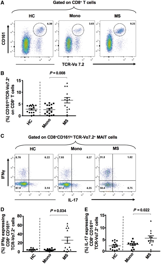
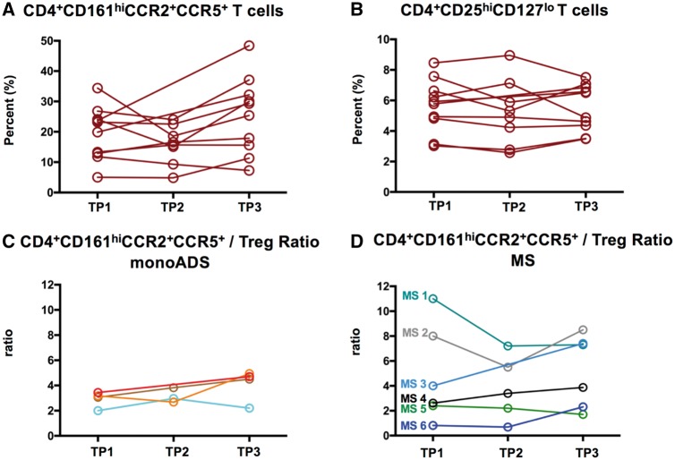
Elucidation of distinct T-cell subsets involved in multiple sclerosis immune-pathophysiology continues to be of considerable interest since an ultimate goal is to more selectively target the aberrant immune response operating in individual patients. While abnormalities of both effector (Teff) and regulatory (Treg) T cells have been reported in patients with multiple sclerosis, prior studies have mostly assessed average abnormalities in either limb of the immune response, rather than both at the same time, which limits the ability to evaluate the balance between effectors and regulators operating in the same patient. Assessing both phenotypic and functional responses of Teffs and Tregs has also proven important. In studies of adults with multiple sclerosis, in whom biological disease onset likely started many years prior to the immune assessments, an added challenge for any reported abnormality is whether the abnormality indeed contributes to the disease (and hence of interest to target therapeutically) or merely develops consequent to inflammatory injury (in which case efforts to develop targeted therapies are unlikely to be beneficial). Paediatric-onset multiple sclerosis, though rare, offers a unique window into early disease mechanisms. Here, we carried out a comprehensive integrated study, simultaneously assessing phenotype and functional responses of both effector and regulatory T cells in the same children with multiple sclerosis, monophasic inflammatory CNS disorders, and healthy controls, recruited as part of the multicentre prospective Canadian Pediatric Demyelinating Disease Study (CPDDS). Stringent standard operating procedures were developed and uniformly applied to procure, process and subsequently analyse peripheral blood cells using rigorously applied multi-parametric flow cytometry panels and miniaturized functional assays validated for use with cryopreserved cells. We found abnormally increased frequencies and exaggerated pro-inflammatory responses of CD8+CD161highTCR-Vα7.2+ MAIT T cells and CD4+CCR2+CCR5+ Teffs in paediatric-onset multiple sclerosis, compared to both control groups. CD4+CD25hiCD127lowFOXP3+ Tregs of children with multiple sclerosis exhibited deficient suppressive capacity, including diminished capacity to suppress disease-implicated Teffs. In turn, the implicated Teffs of multiple sclerosis patients were relatively resistant to suppression by normal Tregs. An abnormal Teff/Treg ratio at the individual child level best distinguished multiple sclerosis children from controls. We implicate abnormalities in both frequencies and functional responses of distinct pro-inflammatory CD4 and CD8 T cell subsets, as well as Treg function, in paediatric-onset multiple sclerosis, and suggest that mechanisms contributing to early multiple sclerosis development differ across individuals, reflecting an excess abnormality in either Teff or Treg limbs of the T cell response, or a combination of lesser abnormalities in both limbs.

Keywords: CD4+CCR2+CCR5+ T cells; CD8+ MAIT cells; multiple sclerosis; paediatric-onset multiple sclerosis; regulatory T cells.


Figures

Figure 1
Figure 1
Increased frequencies and pro-inflammatory cytokine responses of CD8+CD161hi TCR-Vα7.2+ MAIT cells in children with multiple sclerosis. PBMC were stained with appropriate antibodies and isotype controls and initially gated on singlet, live, total CD8+CD3+ T cells (Supplementary Fig. 1). (A) Flow cytometry dot-plots depict approach used to gate on CD8+ CD161hiTCR-Vα7.2+ MAIT cells in representative healthy control (HC), monoADS (Mono) or multiple sclerosis (MS) children. (B) Percentages of circulating MAIT cells in healthy control (n = 16), monoADS (n = 16) and multiple sclerosis (n = 14) children. Two-tailed non-parametric Mann-Whitney U-test for independent groups of monoADS and multiple sclerosis z-scores using the mean and SD of the healthy control group; U = 48, P = 0.008. (C) Following short-term activation with PMA/ionomycin and Golgi stop, PBMC were stained for both surface markers and intracellular-cytokines. Dot plots are shown for representative healthy control, monoADS or multiple sclerosis children, depicting IFNγ and IL-17 expression by activated CD8+CD161hiTCR-Vα7.2+ MAIT cells. (D and E) Percentages of IFNγ and IL-17 expressing MAIT cells in healthy control (n = 14); monoADS (n = 11) and multiple sclerosis (n = 13) children. Mann-Whitney U = 35, P = 0.034 for IFNγ and U = 25.5, P = 0.022 for IL-17.
Figure 2
Figure 2
Increased frequencies and pro-inflammatory cytokine responses of CD4+CCR2+CCR5+ T cells in children with multiple sclerosis. PBMC were stained with appropriate antibodies and isotype controls and initially gated on singlet, live, total CD4+CD3+ T cells (Supplementary Fig. 1). (A) Dot-plots depicting approach to gating on CD4+CCR2+CCR5+ T cells in representative healthy control (HC), monoADS (Mono) or multiple sclerosis (MS) children. (B) Percentages of circulating CD4+CCR2+CCR5+ T cells in healthy control (n = 17), monoADS (n = 18) and multiple sclerosis (n = 15). Two-tailed non-parametric Mann-Whitney U-test for independent groups of monoADS and multiple sclerosis z-scores using the mean and SD of the healthy control; U = 71, P = 0.021. (C) Following short-term activation with PMA/ionomycin and Golgi stop, PBMC were stained for both surface markers and intracellular-cytokines. Dot plots are shown for healthy control, monoADS or multiple sclerosis children depicting IFNγ and IL-17 expression by activated CD4+CCR2+CCR5+ T cells. (D and E) Percentages of IFNγ and IL-17 expressing CD4+CCR2+CCR5+ T cells in healthy control (n = 13), monoADS (n = 13) and multiple sclerosis (n = 14). Mann-Whitney U = 19, P = 0.034 for IFNγ and U = 44, P = 0.023 for IL-17.
Figure 3
Figure 3
Reduced FoxP3 expression and suppressive capacity of circulating CD4+CD25hiCD127low Tregs, and relative resistance to suppression by disease-implicated effector T cells, in children with multiple sclerosis. PBMC were stained with appropriate antibodies and isotype controls and initially gated on singlet, live, total CD4+CD3+ T cells (Supplementary Fig. 1). (A) Dot-plots gating within CD4+CD3+ T cells on CD25hiCD127low T cells as an estimate of circulating Tregs in representative multiple sclerosis, monoADS (Mono) or healthy control (HC) children. (B) Similar Treg frequencies were observed across healthy control (n = 18), monoADS (n = 21) and multiple sclerosis (n = 15) groups. Two-tailed non-parametric Mann-Whitney U-test for independent groups of monoADS and multiple sclerosis z-scores using the mean and SD of the healthy control group; U = 144, P = 0.665. (C) Decreased FOXP3 expression by CD25hiCD127low Tregs of children with multiple sclerosis compared to controls; U = 26, P = 0.041. (D) Tregs of children with multiple sclerosis have reduced capacity to suppress CD4+CD25CD127+ responder (Tresp) cells. Treg suppression assays were performed using FACS-sorted CD4+CD25CD127+ responder (Tresp) cells and CD4+CD25hiCD127low Tregs (Supplementary Fig. 5 for gating strategy). To compare Treg functional capacity across paediatric cohorts, constant numbers of Tresp cells (1.2 × 10³ cells) from healthy control donors were cultured alone or with CD4+CD25hiCD127low Tregs isolated from children with multiple sclerosis, monoADS or healthy controls, at Treg/Tresp ratios of 0:1, 1:8, 1:4 and 1:2; n = 7 children studied in each group). Cells were cultured in triplicates in 96-well U-bottom plates in the presence of plate-bound anti-CD3 (1 µg/ml) and anti-CD28 (1 µg/ml) and incubated for 5 days at 37°C then pulsed 3H-thymidine for an additional 16 h of culture (Supplementary material). Proliferation (counts per minute, cpm) of Tresp cells is shown in the absence or presence of Tregs, at the indicated ratios, using whisker plots (depicting median, interquartile interval, minimum, maximum). Non-parametric Wilcoxon signed rank test was used; Z1:8 = −2.108, P = 0.03; Z1:4 = −1.725, P = 0.08; Z1:2 = −2.492, P = 0.01. (E) Tregs of children with multiple sclerosis have reduced capacity to suppress the disease-implicated CD4+CCR2+CCR5+ Teffs. Constant numbers of FACS-sorted CD4+CCR2+CCR5+ disease-implicated Teffs from healthy control donors were cultured alone or co-cultured with CD4+CD25hiCD127low Tregs from children with multiple sclerosis, monoADS or healthy controls, using the same ratios and assessment of proliferation as described above. Non-parametric Wilcoxon signed rank test; Z1:8 = −1.214, P = 0.22; Z1:4 = −2.492, P = 0.01; Z1:2 = −2.747, P = 0.006. (F) Tresps and Teffs of children with multiple sclerosis are relatively resistant to suppression by normal Tregs. Constant numbers of FACS-sorted CD4+CD25CD127+ Tresp or CD4+CCR2+CCR5+ Teff T cells (1.2 × 10³ cells) from children with multiple sclerosis or healthy controls, were cultured alone or co-cultured with healthy control CD4+CD25hiCD127low Tregs at 0:1, 1:8, 1:4 and 1:2 Treg/Teff ratios. The figure depicts representative results at a single Treg:Teff ratio (1:2). ‘Per cent suppression’ of proliferation was determined as 1 − (cpm incorporated in the co-culture/cpm of responder T cells alone) × 100%. (n = 7). Non-parametric Wilcoxon signed rank test; Z Tres P = −2.625, P = 0.009 and Z Teff = −3.077, P = 0.002.
Figure 4
Figure 4
CCR2+CCR5+ Teff cells are enriched within CD4+CD161+ T cells, particularly in children with multiple sclerosis, who exhibit an abnormal balance between disease-implicated CD4+CD161+CCR2+CCR5+ Teffs and Tregs. PBMC were stained with appropriate antibodies and isotype controls and initially gated on singlet, live, total CD4+CD3+ T cells (Supplementary Fig. 1). (A) Flow cytometry dot-plots depicting approach to gating on CD4+CD161+ T cells in representative healthy control (HC), monoADS (Mono) or multiple sclerosis (MS) children. (B) Percentages of circulating CD4+CD161+ T cells in healthy control (n = 10), monoADS (n = 13) and multiple sclerosis (n = 12). Two-tailed non-parametric Mann-Whitney U-test for independent groups of monoADS and multiple sclerosis z-scores using the mean and SD of the healthy control group; U = 10, P < 0.001. (C) Flow cytometry dot plots depicting co-expression of CCR2 and CCR5 within CD4+CD161+ T cells, in representative healthy control, monoADS, or multiple sclerosis children. (D) Percentages of circulating CCR2+CCR5+ T cells among total CD4+ T cells and among CD4+CD161+ T cells in healthy control (n = 7), monoADS (n = 10) and multiple sclerosis (n = 8); Mann-Whitney U = 13, P = 0.016. (E) The ratio between the frequency of CCR2+CCR5+ T cells (within CD4+CD161+ T cells) and the frequency of Tregs in individual children with multiple sclerosis (n = 8) compared to either monoADS (n = 9) or healthy control (n = 8); Mann-Whitney U = 10, P < 0.001.
Figure 5
Figure 5
The Teff/Treg ratio tends to be a relatively stable characteristic of individuals subjects. Thirty PBMC samples were serially assessed at three time points (TP1, TP2, TP3; separated over at least 2 years) from six children with multiple sclerosis and four children with monoADS who were not exposed to disease modifying therapy or steroids. Frequencies of Teff (CD161+CCR2+CCR5+ CD4+T cells) and Tregs (CD25hiCD127low CD4+ T cells), and Teff/Treg ratios were determined by flow cytometry. As detailed earlier, with all serial samples from the same individual assessed side by side, in the same experiment. (A) Serial measurements of the CD161+CCR2+CCR5+ CD4+ Teff frequencies in both multiple sclerosis and monoADS children; (B) serial measurements of CD4+CD25hiCD127lo Treg frequencies in both multiple sclerosis and monoADS children. (C) Serial Teff/Treg ratios in children with monoADS. (D) Serial Teff/Treg ratios in children with multiple sclerosis. Teff/Treg ratios of the monoADS children tended to be low and relatively stable over time; in children with multiple sclerosis, greater variability in Teff/Treg ratios was observed across individuals, though individual children tended, overall, to maintain their ratio over time.
Figure 6
Figure 6
A normal immune state requires balance between effector and regulatory immune functions. A normal state of immune balance can exist across a broad range of Teff and Treg functions. Imbalances between Teffs and Tregs can manifest as emergence of an autoimmune disease such as multiple sclerosis (when Teff ≫ Treg) or the development of cancer or uncontrolled infection (Teff ≪ Treg).

Comment in

References

    1. Abrahamsson SV, Angelini DF, Dubinsky AN, Morel E, Oh U, Jones JL, et al.Non-myeloablative autologous haematopoietic stem cell transplantation expands regulatory cells and depletes IL-17 producing mucosal-associated invariant T cells in multiple sclerosis. Brain 2013; 136 (Pt 9): 2888–903. - PMC - PubMed
    1. Annibali V, Ristori G, Angelini DF, Serafini B, Mechelli R, Cannoni S, et al.CD161(high)CD8+T cells bear pathogenetic potential in multiple sclerosis. Brain 2011; 134 (Pt 2): 542–54. - PubMed
    1. Balashov KE, Rottman JB, Weiner HL, Hancock WW. CCR5(+) and CXCR3(+) T cells are increased in multiple sclerosis and their ligands MIP-1alpha and IP-10 are expressed in demyelinating brain lesions. Proc Natl Acad Sci USA 1999; 96: 6873–8. - PMC - PubMed
    1. Balint B, Haas J, Schwarz A, Jarius S, Furwentsches A, Engelhardt K, et al.T-cell homeostasis in pediatric multiple sclerosis: old cells in young patients. Neurology 2013; 81: 784–92. - PubMed
    1. Banwell B, Bar-Or A, Arnold DL, Sadovnick D, Narayanan S, McGowan M, et al.Clinical, environmental, and genetic determinants of multiple sclerosis in children with acute demyelination: a prospective national cohort study. Lancet Neurol 2011; 10: 436–45. - PubMed

Publication types

Grants and funding