

doi: 10.1002/mds.27628.
Epub 2019 Feb 13.
Parkinson's disease: Is it a consequence of human brain evolution?
Affiliations
- PMID: 30759321
- PMCID: PMC6593760
- DOI: 10.1002/mds.27628
Item in Clipboard
Parkinson's disease: Is it a consequence of human brain evolution?
Mov Disord.
2019 Apr.
No abstract available
Figures
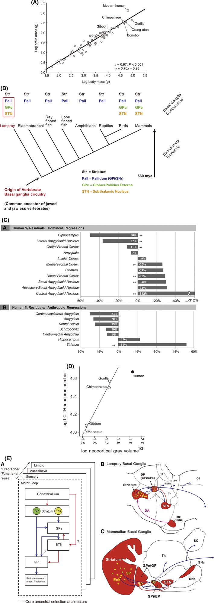
Evolutionary aspects of BG components in humans. (A) Human brain mass larger by scaling plot based on primates40 [with permission]. (B) Clade of the BG: all parts already present in lampreys41 [with permission]. (C) Negative deviations from regression line for striatum and amygdaloid nuclei in humans17 [with permission]. (D) Human LC (vertical axis) shows fewer neurons than expected by comparison to neocortex volume (horizontal axis); TH = tyrosine hydroxylase19 [with permission]. (E) Diagram showing the evolutionarily conserved functional module of the motor loop and subsequent “copy paste” of this module for other functions by the exaptation principle. Blue = GABAergic projections; red = glutamatergic projections; DP = dorsal pallidum; Enk = enkephalin; EP = entopeduncular nucleus; GP = globus pallidus; Gpe = external segment of the globus pallidus; Hb = habenula; ntp = nucleus tuberculi posterior; SP = substance P; Th = thalamus41 [with permission].

Schematic depicting the key features of system proneness to PD pathology. (A) Telencephalization is exponentially larger in humans than in other mammals, here exemplified as rodents. In parallel, the branching of the BG neurons is considerably larger. Slow pacemaking cells are presumably hyperbranching. Their autonomous cell activity provides long‐range neuromodulation and relies on feed‐forward bioenergetics control, with calcium‐dependent and energy‐expensive cellular metabolism, endangering mitochondria, especially at the α‐synuclein–rich synapses. Once a certain threshold is reached, here schematized as a horizontal bar, synaptic and axonal dysfunction occurs, giving rise to first vague and later more concise clinical symptoms. This cascade of events may occur synchronously in different systems with a similar cellular at‐risk phenotype. The slower growth of the BG in comparison to the telencephalon as well as the long human life span promote these processes. (B) PD‐prone systems are characterized by hyperbranching axons regardless of the neurotranmitters.Because hyperbranching neurons project to wide brain areas, related symptoms are multifaceted. Each dot represents neuronal groups with projecting axons indicated by arrows. Of note, nonmotor symptoms transmitted by the dopaminergic VTA are also included, although disease susceptibility is much higher in SNc than VTA neurons, reflecting the distinctive physiology, bioenergetic control mechanisms, and higher numbers of varicosities in the SNc. nbM, nucleus basalis of Meynert; dmX, dorsal motor nucleus of vagus; ggl., ganglia.
References
MeSH terms
Grants and funding
LinkOut - more resources
Full Text Sources
Other Literature Sources
Medical
