

Refining initial diagnosis of Parkinson's disease after follow-up: A 4-year prospective clinical and magnetic resonance imaging study
- PMID: 30759325
- PMCID: PMC6593994
- DOI: 10.1002/mds.27621
Refining initial diagnosis of Parkinson's disease after follow-up: A 4-year prospective clinical and magnetic resonance imaging study
Abstract
Background: No prospective study of patients with Parkinson's disease (PD) has investigated the appearance of vertical gaze abnormalities, a feature suggestive of progressive supranuclear palsy (PSP).
Objective: To identify, within a cohort of patients with an initial diagnosis of PD, those who developed vertical gaze abnormalities during a 4-year follow-up, and to investigate the performance of new imaging biomarkers in predicting vertical gaze abnormalities.
Methods: A total of 110 patients initially classified as PD and 74 controls were enrolled. All patients underwent clinical assessment at baseline and every year up to the end of the follow-up. The pons/midbrain area ratio 2.0 and the Magnetic Resonance Parkinsonism Index 2.0 were calculated.
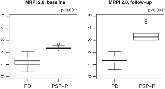
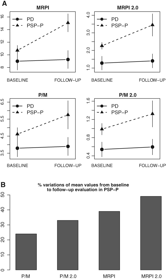
Results: After 4-year follow-up, 100 of 110 patients maintained the diagnosis of PD, whereas 10 PD patients (9.1%) developed vertical gaze abnormalities, suggesting an alternative diagnosis of PSP-parkinsonism. At baseline, the Magnetic Resonance Parkinsonism Index 2.0 was the most accurate biomarker in differentiating PD patients who developed vertical gaze abnormalities from those who maintained an initial diagnosis of PD. At the end of follow-up, both of these biomarkers accurately distinguished PSP-parkinsonism from PD.
Conclusions: Our results demonstrate that a number of patients with an initial diagnosis of PD developed vertical gaze abnormalities during a 4-year follow-up, and the diagnosis was changed from PD to PSP-parkinsonism. In PD patients, baseline Magnetic Resonance Parkinsonism Index 2.0 showed the best performance in predicting the clinical evolution toward a PSP-parkinsonism phenotype, enabling PSP-parkinsonism patients to be identified at the earliest stage of the disease for promising disease-modifying therapies. © 2019 The Authors. Movement Disorders published by Wiley Periodicals, Inc. on behalf of International Parkinson and Movement Disorder Society.
Keywords: magnetic Resonance Parkinsonism Index 2.0; magnetic resonance parkinsonism index; pons/midbrain area ratio 2.0; progressive supranuclear palsy-parkinsonism; vertical gaze abnormalities.
© 2019 The Authors. Movement Disorders published by Wiley Periodicals, Inc. on behalf of International Parkinson and Movement Disorder Society.
Figures
References
-
- Rizzo G, Copetti M, Arcuti S, Martino D, Fontana A, Logroscino G. Accuracy of clinical diagnosis of Parkinson disease: a systematic review and meta‐analysis. Neurology 2016;86:566‐576. - PubMed
-
- Hughes AJ, Daniel SE, Ben‐Shlomo Y, Lees AJ. The accuracy of diagnosis of parkinsonian syndromes in a specialist movement disorder service. Brain 2002;125:861‐870. - PubMed
-
- Joutsa J, Gardberg M, Röyttä M, Kaasinen V. Diagnostic accuracy of parkinsonism syndromes by general neurologists. Parkinsonism Relat Disord 2014;20:840‐844. - PubMed
-
- Caslake R, Moore JN, Gordon JC, Harris CE, Counsell C. Changes in diagnosis with follow‐up in an incident cohort of patients with parkinsonism. J Neurol Neurosurg Psychiatry 2008;79:1202‐1207. - PubMed
MeSH terms
LinkOut - more resources
Full Text Sources
Medical
Miscellaneous
