. 2019 Feb 12;26(7):1681-1690.e5.
doi: 10.1016/j.celrep.2019.01.058.

Structural Basis for Recognition of Ubiquitylated Nucleosome by Dot1L Methyltransferase

Affiliations

Structural Basis for Recognition of Ubiquitylated Nucleosome by Dot1L Methyltransferase

Cathy J Anderson et al. Cell Rep. .

Abstract

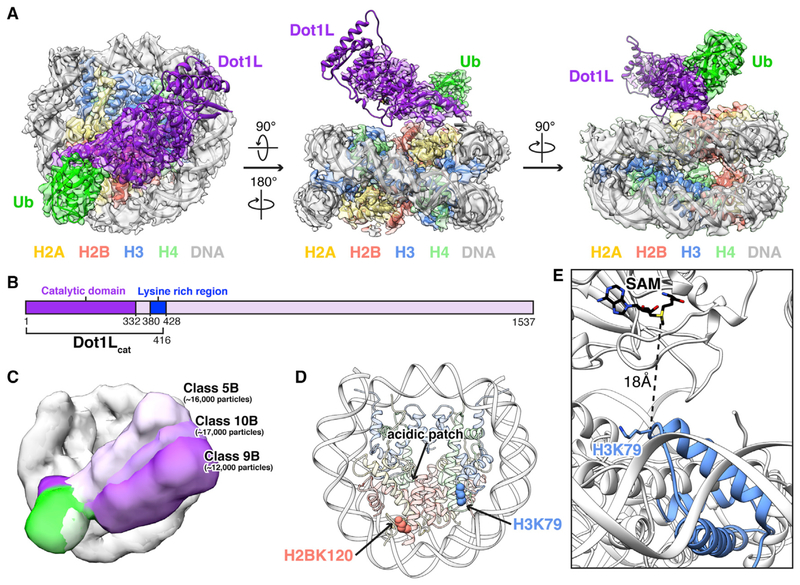
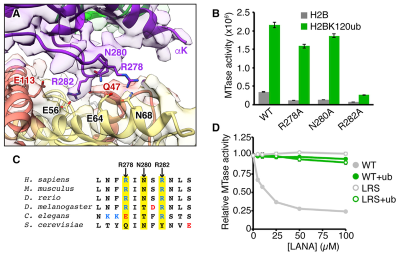
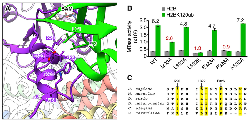
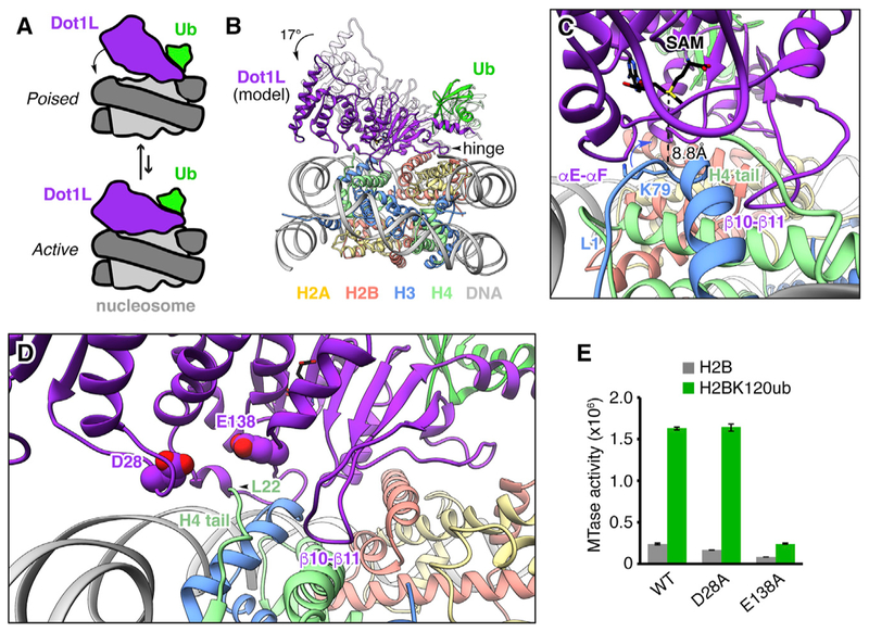
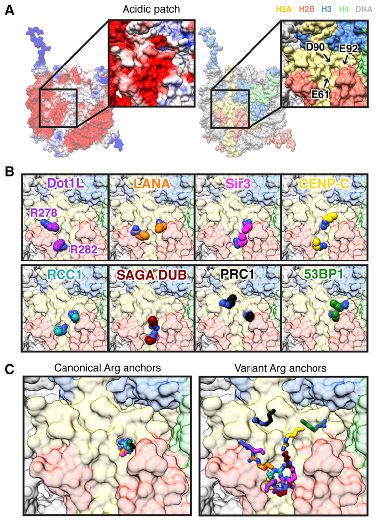
Histone H3 lysine 79 (H3K79) methylation is enriched on actively transcribed genes, and its misregulation is a hallmark of leukemia. Methylation of H3K79, which resides on the structured disk face of the nucleosome, is mediated by the Dot1L methyltransferase. Dot1L activity is part of a trans-histone crosstalk pathway, requiring prior histone H2B ubiquitylation of lysine 120 (H2BK120ub) for optimal activity. However, the molecular details describing both how Dot1L binds to the nucleosome and why Dot1L is activated by H2BK120 ubiquitylation are unknown. Here, we present the cryoelectron microscopy (cryo-EM) structure of Dot1L bound to a nucleosome reconstituted with site-specifically ubiquitylated H2BK120. The structure reveals that Dot1L engages the nucleosome acidic patch using a variant arginine anchor and occupies a conformation poised for methylation. In this conformation, Dot1L and ubiquitin interact directly through complementary hydrophobic surfaces. This study establishes a path to better understand Dot1L function in normal and leukemia cells.

Keywords: Dot1L; chromatin; cryo-EM; histone methyltransferase; nucleosome; single particle; structural biology; ubiquitin.


Conflict of interest statement

DECLARATION OF INTERESTS

The authors declare no competing interests.

Figures

Figure 1.
Figure 1.. Cryo-EM Structure of Dot1L-H2BK120ub Nucleosome Complex
For a Figure360 author presentation of Figure 1, see https://doi.org/10.1016/j.celrep.2019.01.058. (A) Three orthogonal views of the complex with reconstructed map overlaying molecular model. (B) Scheme of human Dot1L highlighting catalytic domain (dark purple), lysine rich region (blue), and minimal Dot1Lcat fragment used in our studies. (C) Overlay of 3D subclasses showing conformational heterogeneity of Dot1L (shades of purple) and ubiquitin (shades of green). (D) Nucleosome structure with acidic patch and sites of ubiquitylation (H2BK120) and Dot1L-mediated methylation (H3K79) indicated. (E) Zoomed view of Dot1L active site and targeted lysine in our model with distance between H3K79 Ca and SAM cofactor sulfur indicated. See also Figures S1–S4 and Tables S1 and S2.
Figure 2.
Figure 2.. Dot1L Interacts with the Nucleo-some Acidic Patch
(A) Zoomed view of Dot1L nucleosome-interaction loop engaged with nucleosome acidic patch surface. Model, reconstructed map, and relevant side chains are shown. (B) Quantified Dot1L methyltransferase (MTase) assay using indicated mutants with unmodified (H2B) and ubiquitylated (H2BK120ub) nucleosomes. (C) Sequence alignment of Dot1L nucleosome-interaction loop with positively and negatively charged residues colored blue and red, respectively, and key positions highlighted in yellow. (D) Competitive MTase assay with varying concentrations of wild-type (WT) or nucleosome-binding-deficient (LRS = LRS 8–10 AAA) LANA fusion proteins on unmodified and H2BK120ub nucleosomes. Five replicates performed for all assays and means and SDs are shown. See also Figure S5.
Figure 3.
Figure 3.. Ubiquitin Interacts with Dot1L to Enhance H3K79 Methylation
(A) Zoomed view of Dot1L-ubiquitin interaction with relevant side chains indicated. (B) Quantified MTase assay of Dot1L mutants on unmodified (H2B) and ubiquitylated (H2BK120ub) nucleosomes. Numbers above bars indicate fold enhancement by ubiquitin for each Dot1L protein. A value of 1 indicates no difference in activity toward H2BK120ub relative to unmodified nucleosomes. Five replicates performed for all assays and means and SDs are shown. (C) Sequence alignment of Dot1L ubiquitin-binding regions with key positions highlighted in yellow. See also Figure S6.
Figure 4.
Figure 4.. Model of Active Conformation of Dot1L on the Nucleosome
(A) Scheme of proposed poised to active conformational rearrangement of Dot1L and ubiquitin on the nucleosome. (B) Front view of model of Dot1L and ubiquitin in an active conformation made by a 17° rotation of Dot1L about hinge Arg282 Cα atom of Dot1L. Ubiquitin was adjusted with Dot1L, keeping the orientation of ubiquitin relative to Dot1L constant. Cryo-EM model shown in white for comparison. (C) Zoomed side view of active model with SAM sulfur-H3K79 Cα distance labeled. Loops with anticipated conformational changes are indicated. (D) Front view of active model with key residues of acidic cleft indicated. (E) Quantified MTase assay of Dot1L acidic cleft mutants on unmodified (H2B) and ubiquitylated (H2BK120ub) nucleosomes. Five replicates performed for all assays and means and SDs are shown.
Figure 5.
Figure 5.. Canonical and Variant Arginine Anchors Bind the Nucleo-some Acidic Patch
(A) Electrostatic (generated with Adaptive Poisson-Boltzmann Solver [APBS] using AMBER) and histone-colored surfaces of nucleosome. (B) Zoomed views of arginine anchors of Dot1L, LANA (PDB: 1ZLA), Sir3 (PDB: 3TU4), CENP-C (PDB: 4X23), RCC1 (PDB: 3MVD), SAGA DUB module (PDB: 4ZUX), PRC1/RING1B (PDB: 4RP8), and 53BP1 (PDB: 5KGF). (C) Overlay of all canonical (left) and variant (right) arginine anchors.

References

    1. Afonine PV, Poon BK, Read RJ, Sobolev OV, Terwilliger TC, Urzhumtsev A, and Adams PD (2018). Real-space refinement in PHENIX for cryo-EM and crystallography. Acta Crystallogr. D Struct. Biol 74, 531–544. - PMC - PubMed
    1. Armache KJ, Garlick JD, Canzio D, Narlikar GJ, and Kingston RE (2011). Structural basis of silencing: Sir3 BAH domain in complex with a nucleosome at 3.0 Å resolution. Science 334, 977–982. - PMC - PubMed
    1. Barbera AJ, Chodaparambil JV, Kelley-Clarke B, Joukov V, Walter JC, Luger K, and Kaye KM (2006). The nucleosomal surface as a docking station for Kaposi’s sarcoma herpesvirus LANA. Science 311, 856–861. - PubMed
    1. Bernt KM, Zhu N, Sinha AU, Vempati S, Faber J, Krivtsov AV, Feng Z, Punt N, Daigle A, Bullinger L, et al. (2011). MLL-rearranged leukemia is dependent on aberrant H3K79 methylation by DOT1L. Cancer Cell 20, 66–78. - PMC - PubMed
    1. Bilokapic S, Strauss M, and Halic M (2018). Histone octamer rearranges to adapt to DNA unwrapping. Nat. Struct. Mol. Biol 25, 101–108. - PMC - PubMed

Publication types

LinkOut - more resources