Review
. 2019 Apr 26;11(3):032002.
doi: 10.1088/1758-5090/ab06ea.

Electrobiofabrication: electrically based fabrication with biologically derived materials

Affiliations
Review

Electrobiofabrication: electrically based fabrication with biologically derived materials

Jinyang Li et al. Biofabrication. .

Abstract

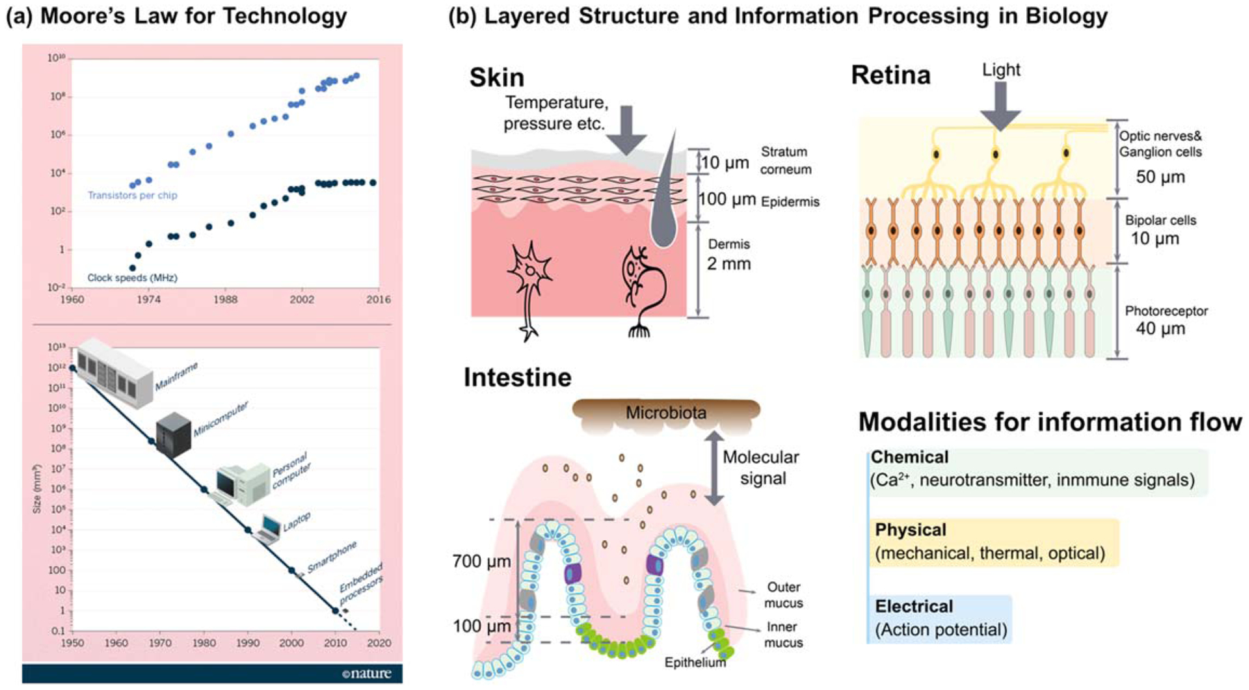
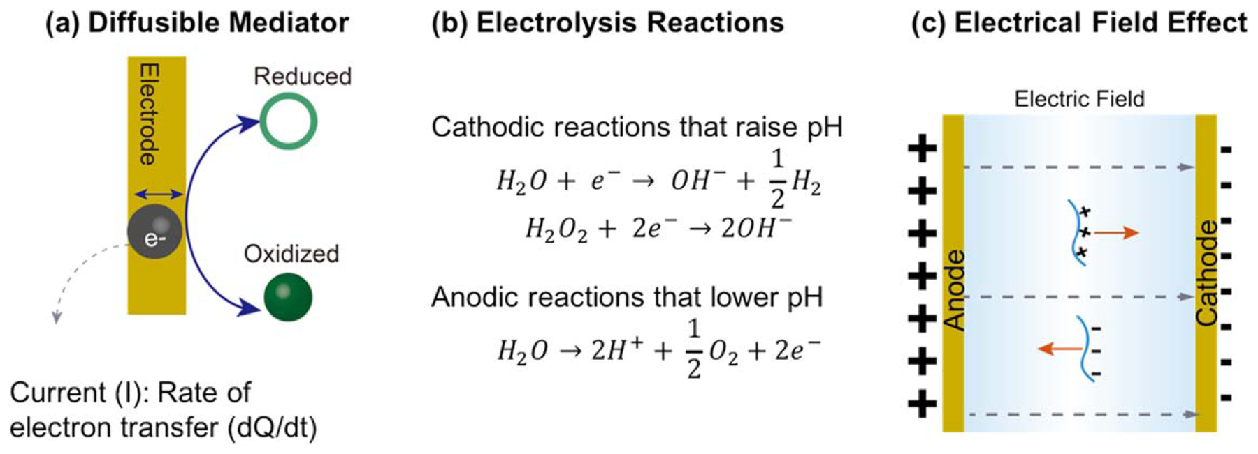
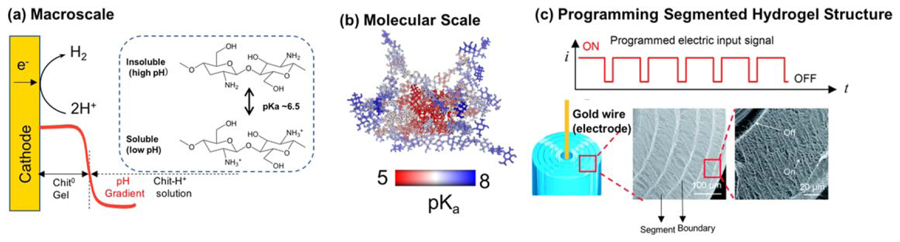
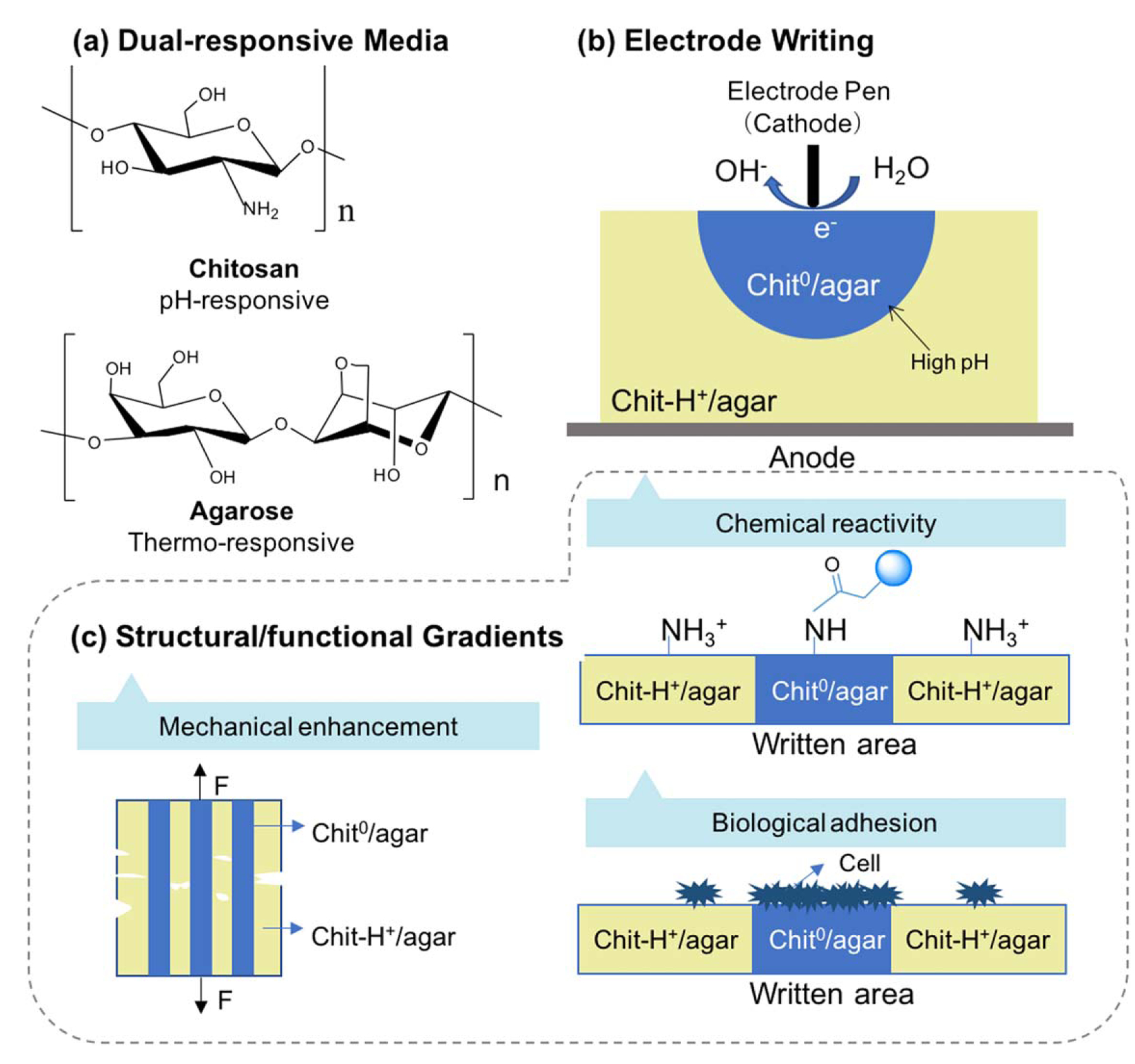
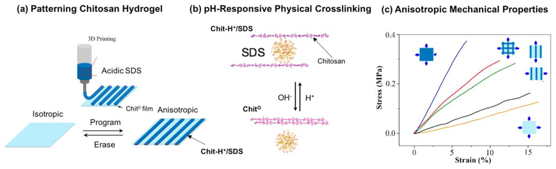
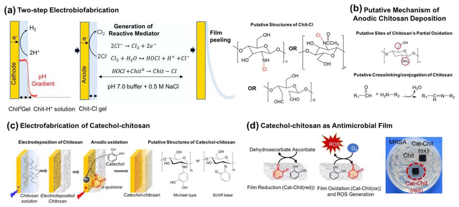
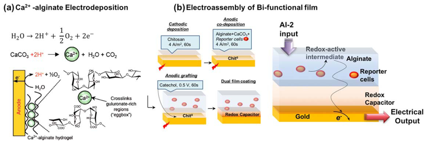
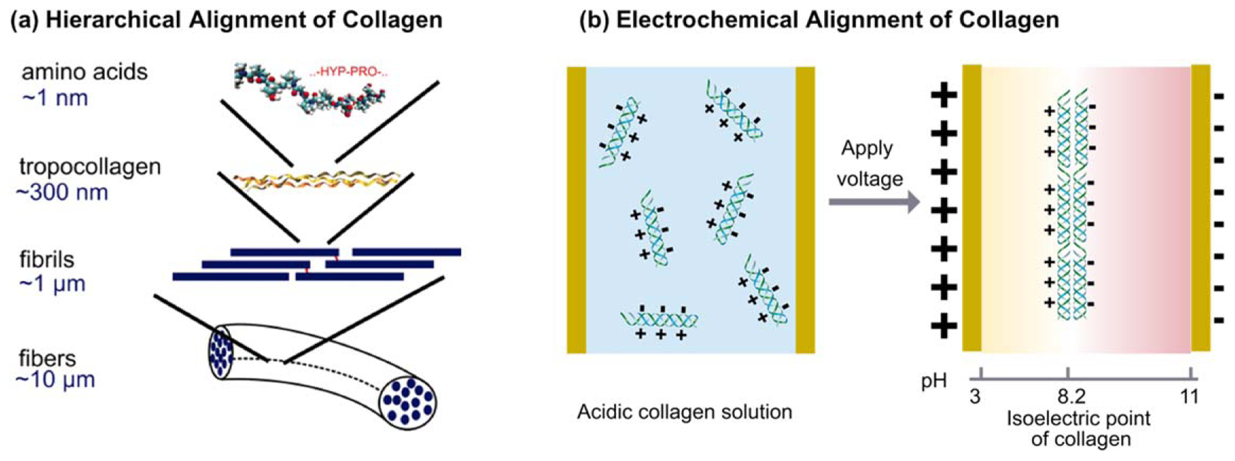
While conventional material fabrication methods focus on form and strength to achieve function, the fabrication of material systems for emerging life science applications will need to satisfy a more subtle set of requirements. A common goal for biofabrication is to recapitulate complex biological contexts (e.g. tissue) for applications that range from animal-on-a-chip to regenerative medicine. In these cases, the material systems will need to: (i) present appropriate surface functionalities over a hierarchy of length scales (e.g. molecular features that enable cell adhesion and topographical features that guide differentiation); (ii) provide a suite of mechanobiological cues that promote the emergence of native-like tissue form and function; and (iii) organize structure to control cellular ingress and molecular transport, to enable the development of an interconnected cellular community that is engaged in cell signaling. And these requirements are not likely to be static but will vary over time and space, which will require capabilities of the material systems to dynamically respond, adapt, heal and reconfigure. Here, we review recent advances in the use of electrically based fabrication methods to build material systems from biological macromolecules (e.g. chitosan, alginate, collagen and silk). Electrical signals are especially convenient for fabrication because they can be controllably imposed to promote the electrophoresis, alignment, self-assembly and functionalization of macromolecules to generate hierarchically organized material systems. Importantly, this electrically based fabrication with biologically derived materials (i.e. electrobiofabrication) is complementary to existing methods (photolithographic and printing), and enables access to the biotechnology toolbox (e.g. enzymatic-assembly and protein engineering, and gene expression) to offer exquisite control of structure and function. We envision that electrobiofabrication will emerge as an important platform technology for organizing soft matter into dynamic material systems that mimic biology's complexity of structure and versatility of function.


Figures

Figure 1.
Figure 1.
Layered thin films are used to process information in technology and biology. (a) Moore’s law tracks progress in information technology using generic metrics for structure and performance. Reprinted by permission from Macmillan Publishers Ltd: [Nature] [6], copyright 2016. (b) A common goal for biofabrication is to recapitulate biology’s layered structures and spatiotemporal signaling.
Figure 2.
Figure 2.
Electrodes provide electrical signals for electrobiofabrication. (a) The current quantifies the electrochemical reactions (i.e. the exchange of electrons at the electrode–solution interface) that can (b) provide localized chemical gradients (e.g. in pH) that cue self-assembly. (c) The electric field can induce macromolecules to migrate and undergo changes in conformation and alignment.
Figure 3.
Figure 3.
Cathodic electrodeposition of chitosan. (a) Chitosan deposition (i.e. self-assembly) is induced by the high pH generated by cathodic electrolysis reactions. (b) Molecular modeling shows the pKa of an individual glucosamine residue is lowered when it is buried within the crystalline domains that serve as the physical network junctions (i.e. crosslinks). Reproduced with permission from [204]. John Wiley & Sons. © 2018 WILEY-VCH Verlag GmbH & Co. KGaA, Weinheim. (c) When an oscillating electrical input sequence is used to cue chitosan’s electrodeposition, the resulting hydrogel has a segmented internal structure that is controlled by the input sequence. Reproduced from [203] with permission from The Royal Society of Chemistry.
Figure 4.
Figure 4.
Cathodic writing onto a dual responsive (chitosan–agarose) medium induces the formation of an internal structure. (a) The dual responsive hydrogel is formed by cooling an acidic blend of chitosan and agarose. (b) The internal structure is created using a cathodic ‘pen’ to create regions of high pH that induce the localized self-assembly of chitosan chains within the agarose network. (c) The gradients in the structure induced by writing also yield gradients in mechanical, chemical and biological properties. Adapted with permission from [204]. John Wiley & Sons. © 2018 WILEY-VCH Verlag GmbH & Co. KGaA, Weinheim.
Figure 5.
Figure 5.
Chitosan can be crosslinked by independent physical mechanisms enabling films to be reversibly patterned, erased and reconfigured. (a) An acidic SDS solution is used as an ‘ink’ that is printed onto a cathodically deposited chitosan film. (b) One physical crosslinking mechanism involves electrostatic interactions between protonated chitosan chains and SDS micelles (Chit-H+-SDS), while a second physical crosslinking mechanism involves the crystalline domains that serve as network junctions (Chit0). (c) Films that are patterned with different crosslinking mechanisms offer anisotropic mechanical properties. Adapted with permission from [42]. John Wiley & Sons. © 2017 WILEY-VCH Verlag GmbH & Co. KGaA, Weinheim.
Figure 6.
Figure 6.
Anodic (oxidative) deposition of chitosan involves covalent modifications. (a) Two-step fabrication of chitosan film to obtain chloramine residues that confer antimicrobial activities. (b) An analogous single step anodic deposition mechanism for chitosan. (c) The anodic fabrication of a catechol–chitosan film. Adapted from [239]. CC BY 4.0. (d) Catechol–chitosan films are redox-active and allow for the sustained in situ generation of ROS that can inhibit the growth of methicillin resistant Staphylococcus aureus (MRSA). Adapted from [240]. Copyright 2018, with permission from Elsevier.
Figure 7.
Figure 7.
Electrobiofabrication of functionalized alginate hydrogel films. (a) The mechanism for the anodic deposition of Ca2+-alginate hydrogels. Reproduced with permission from [143]. John Wiley & Sons. Copyright © 2009 WILEY-VCH Verlag GmbH & Co. KGaA, Weinheim. (b) Three-step electrobiofabrication of a dual functional film that transduces the detection of a bacterial signaling molecule (autoinducer-2; AI-2) into a redox-active intermediate that is detected electrochemically. Adapted with permission from [266]. John Wiley & Sons. © 2016 WILEY-VCH Verlag GmbH & Co. KGaA, Weinheim.
Figure 8.
Figure 8.
Electrical signals can be used to induce the migration, alignment and assembly of proteins. (a) The hierarchical organization of collagen. Reproduced with permission from [272]. © 2006 by The National Academy of Sciences of the USA. (b) Electrochemical alignment of collagen at a location where the pH is equal to its isoelectric point.

References

    1. Groll J. et al. Biofabrication: reappraising the definition of an evolving field. Biofabrication. 2016;8:013001. - PubMed
    1. McHugh KJ et al. 2017. Fabrication of fillable microparticles and other complex 3D microstructures Science 357 1138–42 - PMC - PubMed
    1. Gordonov T, Kim E, Cheng Y, Ben-Yoav H, Ghodssi R, Rubloff G, Yin J-J, Payne GF and Bentley WE 2014. Electronic modulation of biochemical signal generation Nat. Nanotechnol 9 605–10 - PubMed
    1. Wang H, Ma X and Hao Y 2017. Electronic devices for human-machine interfaces Adv. Mater. Interfaces 4 1–20
    1. Guo S-Z, Qiu K, Meng F, Park SH and McAlpine MC 2017. 3D printed stretchable tactile sensors Adv. Mater 29 1701218 - PMC - PubMed

Publication types