. 2019 Feb 12;20(3):777.
doi: 10.3390/ijms20030777.

Preclinical Evaluation of Long-Term Neuroprotective Effects of BDNF-Engineered Mesenchymal Stromal Cells as Intravitreal Therapy for Chronic Retinal Degeneration in Rd6 Mutant Mice

Affiliations

Preclinical Evaluation of Long-Term Neuroprotective Effects of BDNF-Engineered Mesenchymal Stromal Cells as Intravitreal Therapy for Chronic Retinal Degeneration in Rd6 Mutant Mice

Renata Lejkowska et al. Int J Mol Sci. .

Abstract

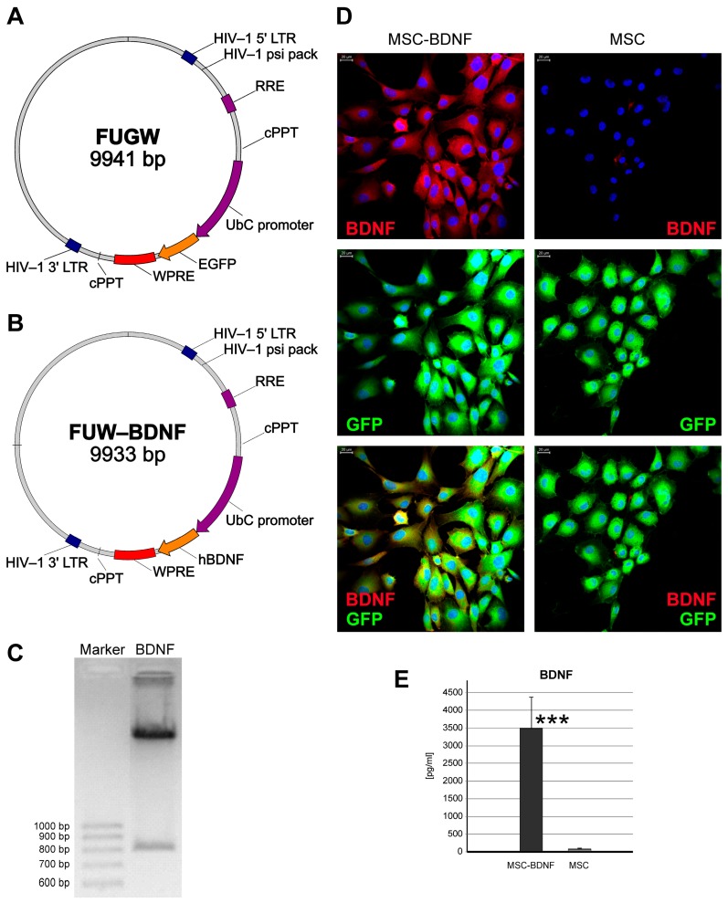
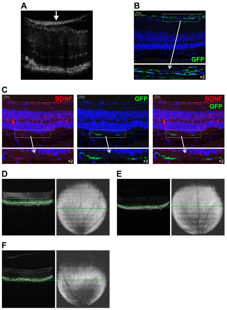
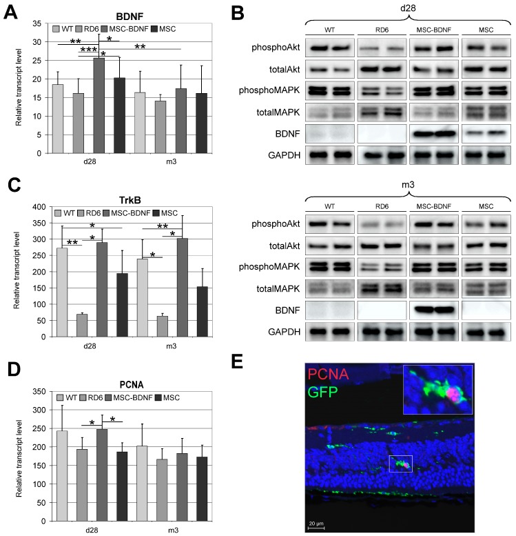
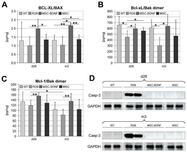
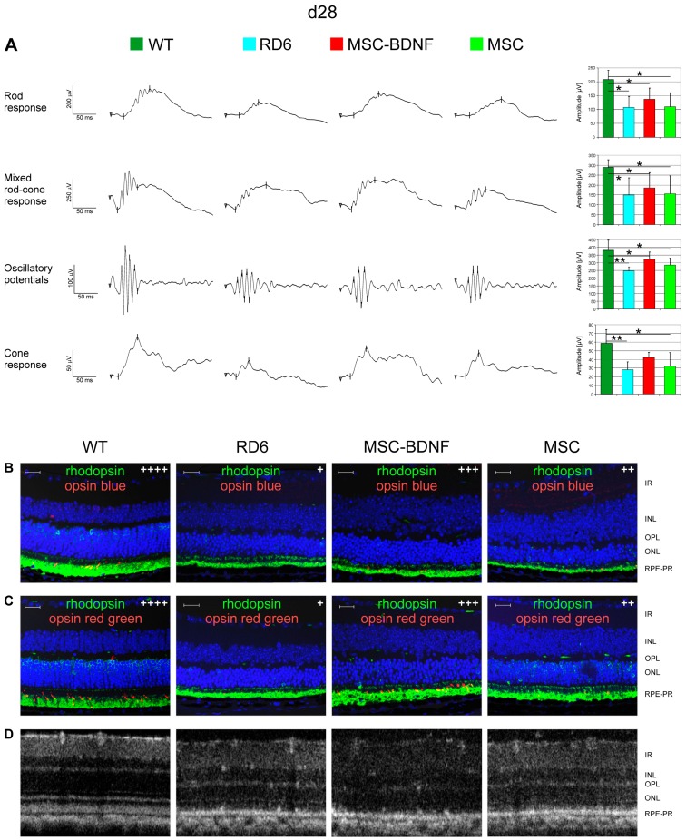
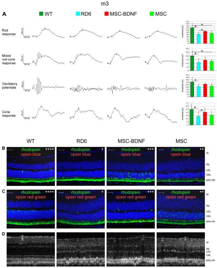
This study aimed to investigate whether the transplantation of genetically engineered bone marrow-derived mesenchymal stromal cells (MSCs) to overexpress brain-derived neurotrophic factor (BDNF) could rescue the chronic degenerative process of slow retinal degeneration in the rd6 (retinal degeneration 6) mouse model and sought to identify the potential underlying mechanisms. Rd6 mice were subjected to the intravitreal injection of lentivirally modified MSC-BDNF or unmodified MSC or saline. In vivo morphology, electrophysiological retinal function (ERG), and the expression of apoptosis-related genes, as well as BDNF and its receptor (TrkB), were assessed in retinas collected at 28 days and three months after transplantation. We observed that cells survived for at least three months after transplantation. MSC-BDNF preferentially integrated into the outer retinal layers and considerably rescued damaged retinal cells, as evaluated by ERG and immunofluorescence staining. Additionally, compared with controls, the therapy with MSC-BDNF was associated with the induction of molecular changes related to anti-apoptotic signaling. In conclusion, BDNF overexpression observed in retinas after MSC-BDNF treatment could enhance the neuroprotective properties of transplanted autologous MSCs alone in the chronically degenerated retina. This research provides evidence for the long-term efficacy of genetically-modified MSC and may represent a strategy for treating various forms of degenerative retinopathies in the future.

Keywords: BDNF; MSC; OCT; lentiviral vectors; rd6; regenerative medicine; retinal degeneration; tissue imaging.


Conflict of interest statement

The authors declare no conflict of interest. The funders had no role in the design of the study; in the collection, analyses, or interpretation of data; in the writing of the manuscript, or in the decision to publish the results.

Figures

Figure 1
Figure 1
Characterization of lentiviral MSCs transduction efficiency. The schemes of plasmids used for lentivirus production for subsequent murine MSCs transduction are shown. The lentiviral backbone plasmid (FUGW) contained the green fluorescent protein (GFP) coding sequence (A) that was removed to insert the human BDNF sequence and then FUGW-BDNF plasmid was created (B) for relevant lentiviral vectors production. The correct band for BDNF insert (765 bp) was observed under ultraviolet (UV) light in agarose gel (C). Quantitative analysis of BDNF levels from MSC-BDNF and unmodified MSC cultures in vitro (D). Noninfected control MSCs produced only trace amount of BDNF, whereas production of BDNF in MSC-BDNF culture was approximately 35-fold increased. These data were corroborated by double immunofluorescent staining of BDNF and GFP proteins for their qualitative expression and co-expression analysis (E). Scale bar: 20 µm, *** p < 0.001.
Figure 2
Figure 2
Long-term follow-up of genetically modified MSC-BDNF and MSC trafficking and homing at different time points post-intravitreal transplantation in rd6 mice. A representative SD-OCT image of chronically degenerated retina of rd6 mouse at the 28th day after intravitreal MSC-BDNF injection (A). A hyperreflective streak of the accumulated MSC (white arrow) at the vitreoretinal interface is observed. A representative fluorescence image of degenerated retina of rd6 mouse at 28 days after intravitreal MSC injection (B). At this time point, the vast majority of the injected GFP-positive cells (green) were found to be located at the vitreoretinal interface and in the superficial ganglion cell layer. A representative fluorescence images of degenerated retina of rd6 mouse at three months after intravitreal MSC-BDNF injection (C). At this time of the experiment, the injected GFP-positive cells (green) were found to be aligned along the RPE-photoreceptor junction and showed double immunostaining against BDNF (red). A representative retinal volume intensity projections of OCT scans of rd6 control mouse (D), after intravitreal MSC-BDNF injection (E) and MSC alone transplantation (F) at the third month of the experiment. At this time of the experiment, the considerable reduction of the retinal white spots that correspond to macrophages and monocytes at the level of retinal pigment epithelium was observed only in eyes after intravitreal MSC-BDNF injection. Green lines indicate the retinal level where the volume intensity projection image (VIP) was captured. Scale bar: 20 µm.
Figure 3
Figure 3
Long-term follow-up of BDNF production and its biological function at different time points post-intravitreal MSC-BDNF transplantation. BDNF mRNA (A), BDNF, phosphoAkt, totalAkt, phosphoMAPK and totalMAPK protein (B) expression was detected in retinas from eyes treated with MSC-BDNF, and their levels were significantly increased post-transplantation compared to other groups: at 28 days in case of mRNA and protein and at three months in the case of BDNF protein only. We also observed increased TrkB gene expression in retinas at 28 days and three months after MSC-BDNF and MSC alone transplantation compared to rd6 and wild type (WT) (C). The follow-up of retinal cell proliferation at different time points post-intravitreal transplantation in rd6 mice was also performed. Quantitative analysis of proliferating cell nuclear antigen (PCNA) mRNA expression revealed that their levels were significantly increased in retinas from eyes treated with MSC-BDNF at 28 days post transplantation compared with those in eyes treated with the PBS and MSCs alone (D). Double-stained sections for PCNA and GFP (endogenous marker of transplanted MSC) used to visualize and localize proliferating cells revealed the extraordinary PCNA protein concentration in MSC-BDNF transplanted 28 days previously (E). Representative images of the performed analyses are shown. Scale bar: 20 µm. Reference gene used for qRT-PCR analysis was glyceraldehyde 3-phospate dehydrogenase (GAPDH). Mean values ± SD are presented in the diagrams, * p < 0.05, ** p < 0.01, *** p < 0.001 (n = 7/group/time point).
Figure 4
Figure 4
The expression profile of selected apoptosis-related molecules in retinas treated with MSC-BDNF or MSC alone at different time points (at 28 days and three months post transplantation) and compared to WT and rd6 mice. The mRNA expression of Bcl-xL and BAX genes was determined by the quantitative PCR and the relative ratio Bcl-xL/BAX was calculated (A). The concentration of Bcl-xL and Bak protein dimer was determined by specific Luminex (B) Similarly, the concentration of Mcl-1/Bak dimer protein was measured (C). Caspase-3 protein expression was determined by Western blot, which revealed a lack of expression of this form in WT mice and rd6 after cell transplantation compared to rd6 mice with no treatment (D). GAPDH served as loading. A representative image is shown. Mean values ± SDs are presented in the diagrams, * p < 0.05, ** p < 0.01 (n = 7/group/time point).
Figure 5
Figure 5
Effects of in vivo experimental therapy with genetically transduced MSC-BDNF and unmodified MSC on the retinal function and OCT-based retinal morphology at 28 days post transplantation compared to rd6 mice with no treatment and WT mice. The representative ERG responses recorded after cell injection are shown (A). The b-wave amplitude measurements are presented as the mean ± SD immunohistofluorescence co-staining for rhodopsin (green) with opsin blue (red) in (B) and rhodopsin (green) with opsin red/green (red) in (C) are displayed in the representative retinal specimens from all the groups with semi quantitative evaluation (signal intensity was marked with subsequent methodology: “+” when less than 5 immunopositive cells present in the image, “++” for 6–15 of visible positive cells, “+++” for 16–30 of positive stained cells, “++++” more than 30 immunopositive cells. The control rd6 mouse retinas (left column) are almost completely negative for both cone opsins, whereas MSC-BDNF-treated retinas displayed regeneration of cones as evidenced by positive immunoreactivity for red/green cone opsin (middle column). The representative OCT images of the retinal sections with their typical layered structure are presented in (D). Retinal layers were marked both at histological images and OCT as follows: IR- inner retina, INL- inner nuclear layer, OPL- outer plexiform layer, ONL- outer nuclear layer, RPE-PR–retinal pigment epithelium and photoreceptors. Scale bar: 20 µm. * p < 0.05; ** p < 0.01.
Figure 6
Figure 6
Effects of in vivo experimental therapy with genetically transduced MSC-BDNF and unmodified MSC on the retinal function and OCT-based retinal morphology at three months post transplantation compared to rd6 mice with no treatment and WT mice. The representative ERG responses recorded after cell injection are shown (A). The b-wave amplitude measurements are presented as the mean ± SD. Immunohistofluorescence co-staining for rhodopsin (green) with opsin blue (red) in (B) and rhodopsin (green) with opsin red/green (red) in (C) are displayed in the representative retinal specimens from all the groups with semi quantitative evaluation (signal intensity was marked with subsequent methodology: “+” when less than 5 immunopositive cells present in the image, “++” for 6–15 of visible positive cells, “+++” for 16–30 of positive stained cells, “++++” more than 30 immunopositive cells. The control rd6 mouse retinas (left column) are almost completely negative for both cone opsins, whereas MSC-BDNF-treated retinas displayed a regeneration of cones as evidenced by the positive immunoreactivity for red/green cone opsin (middle column). The representative OCT images of the retinal sections with their typical layered structure are presented in (D). Retinal layers were marked both at histological images and OCT as follows: IR- inner retina, INL- inner nuclear layer, OPL- outer plexiform layer, ONL- outer nuclear layer, RPE-PR–retinal pigment epithelium and photoreceptors. Scale bar: 20 µm. * p < 0.05.

Similar articles

Cited by

References

    1. Chichagova V., Hallam D., Collin J., Zerti D., Dorgau B., Felemban M., Lako M., Steel D.H. Cellular regeneration strategies for macular degeneration: Past, present and future. Eye (London) 2018;32:946–971. doi: 10.1038/s41433-018-0061-z. - DOI - PMC - PubMed
    1. Park S.S., Moisseiev E., Bauer G., Anderson J.D., Grant M.B., Zam A., Zawadzki R.J., Werner J.S., Nolta J.A. Advances in bone marrow stem cell therapy for retinal dysfunction. Prog. Retin. Eye Res. 2017;56:148–165. doi: 10.1016/j.preteyeres.2016.10.002. - DOI - PMC - PubMed
    1. Jones M.K., Lu B., Girman S., Wang S. Cell-based therapeutic strategies for replacement and preservation in retinal degenerative diseases. Prog. Retin. Eye Res. 2017;58:1–27. doi: 10.1016/j.preteyeres.2017.01.004. - DOI - PMC - PubMed
    1. Pius-Sadowska E., Machalinski B. BDNF—A key player in cardiovascular system. J. Mol. Cell Cardiol. 2017;110:54–60. doi: 10.1016/j.yjmcc.2017.07.007. - DOI - PubMed
    1. Minnone G., De Benedetti F., Bracci-Laudiero L. NGF and Its Receptors in the Regulation of Inflammatory Response. Int. J. Mol. Sci. 2017;18 doi: 10.3390/ijms18051028. - DOI - PMC - PubMed

MeSH terms

LinkOut - more resources