. 2019 Feb 12;11(2):210.
doi: 10.3390/cancers11020210.

TARBP2-Enhanced Resistance during Tamoxifen Treatment in Breast Cancer

Affiliations

TARBP2-Enhanced Resistance during Tamoxifen Treatment in Breast Cancer

Ming-Yang Wang et al. Cancers (Basel). .

Abstract

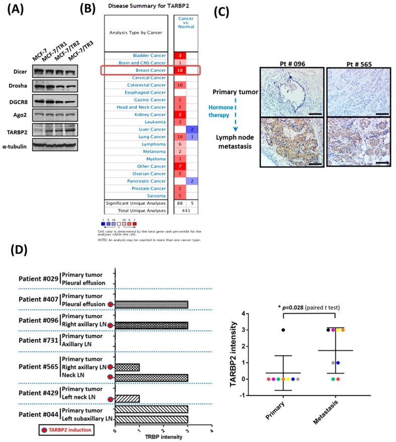
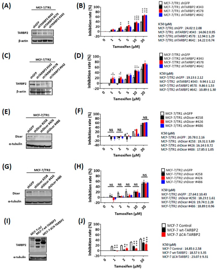
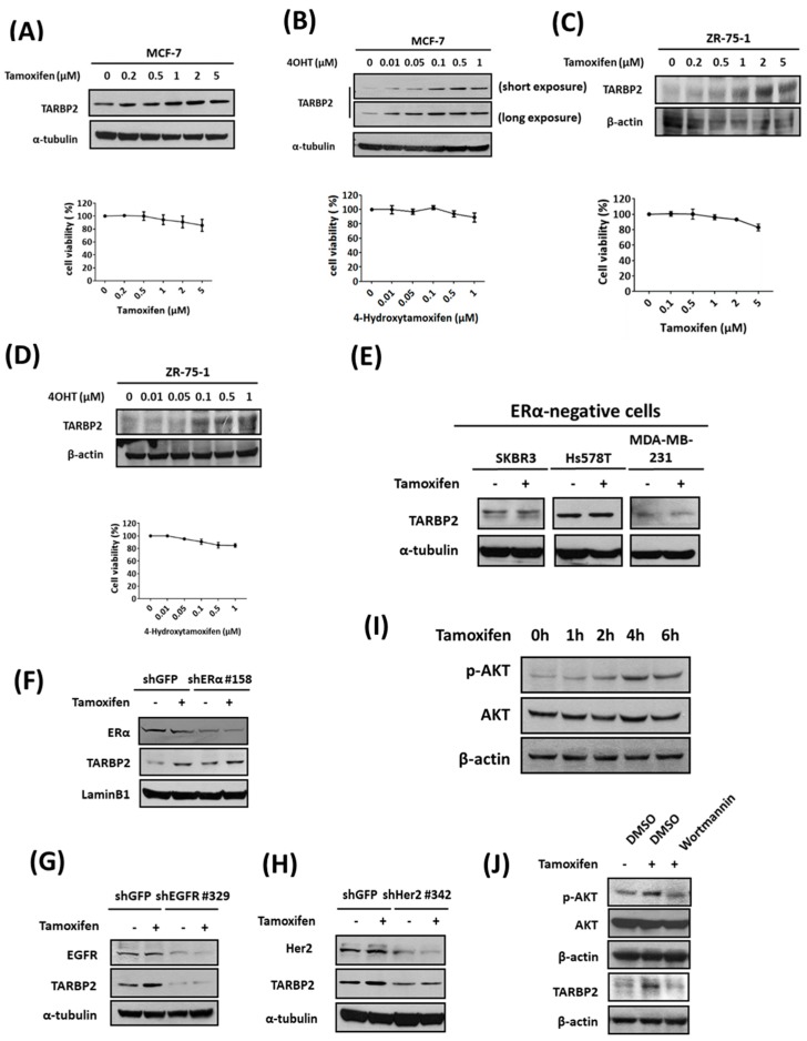
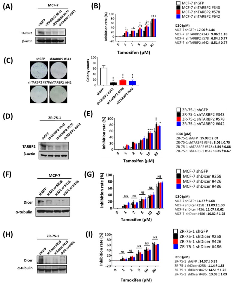
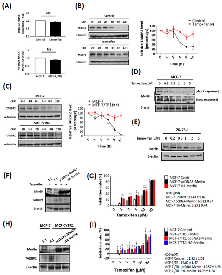
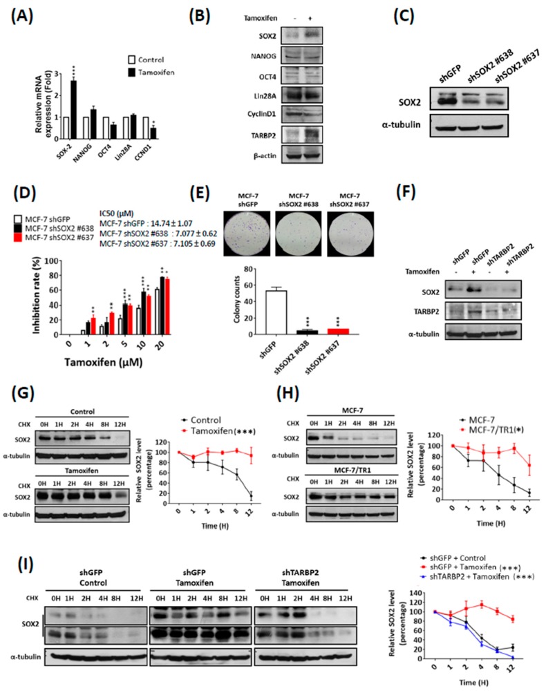
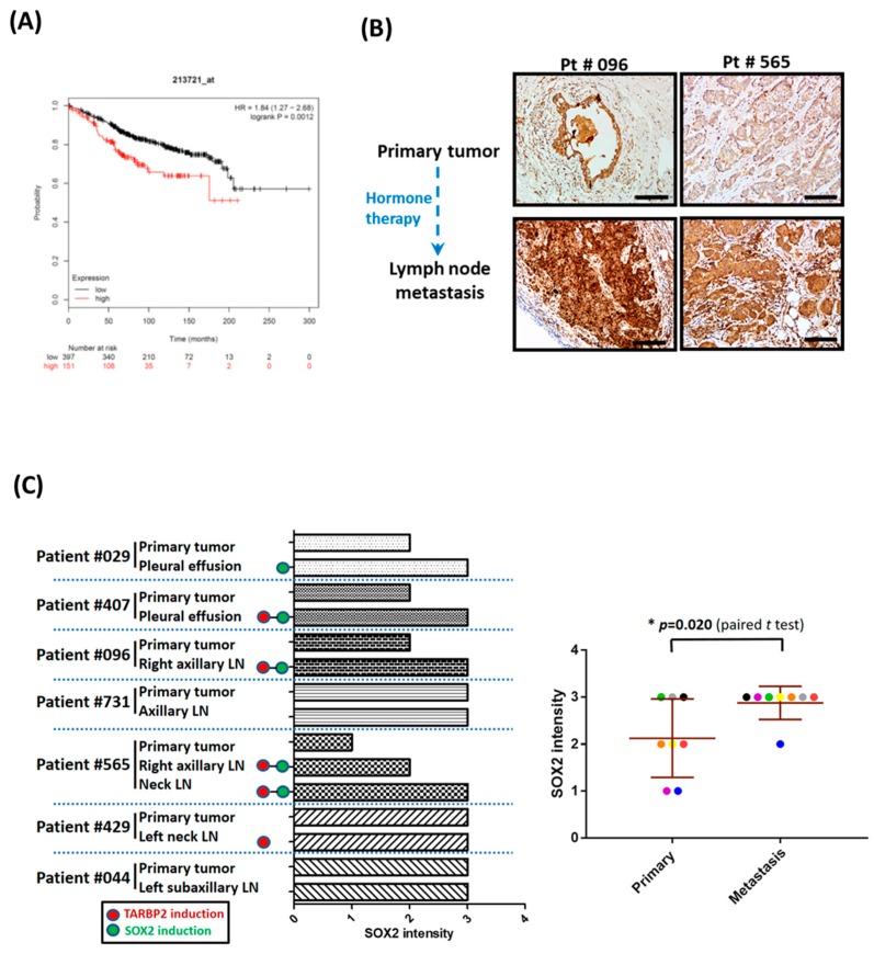
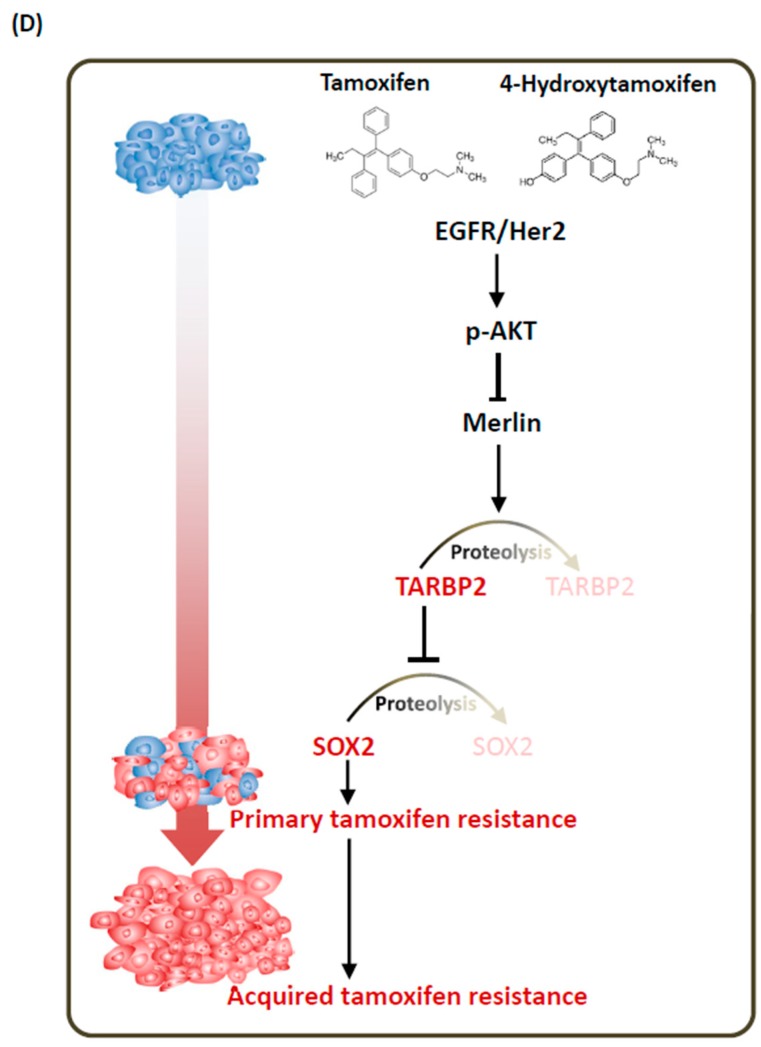
Tamoxifen is the most widely used hormone therapy in estrogen receptor-positive (ER+) breast cancer, which accounts for approximately 70% of all breast cancers. Although patients who receive tamoxifen therapy benefit with respect to an improved overall prognosis, resistance and cancer recurrence still occur and remain important clinical challenges. A recent study identified TAR (HIV-1) RNA binding protein 2 (TARBP2) as an oncogene that promotes breast cancer metastasis. In this study, we showed that TARBP2 is overexpressed in hormone therapy-resistant cells and breast cancer tissues, where it enhances tamoxifen resistance. Tamoxifen-induced TARBP2 expression results in the desensitization of ER+ breast cancer cells. Mechanistically, tamoxifen post-transcriptionally stabilizes TARBP2 protein through the downregulation of Merlin, a TARBP2-interacting protein known to enhance its proteasomal degradation. Tamoxifen-induced TARBP2 further stabilizes SOX2 protein to enhance desensitization of breast cancer cells to tamoxifen, while similar to TARBP2, its induction in cancer cells was also observed in metastatic tumor cells. Our results indicate that the TARBP2-SOX2 pathway is upregulated by tamoxifen-mediated Merlin downregulation, which subsequently induces tamoxifen resistance in ER+ breast cancer.

Keywords: SOX2; TARBP2; hormone therapy; merlin; tamoxifen.


Conflict of interest statement

The authors declare no conflict of interest.

Figures

Figure 1
Figure 1
TARBP2 is overexpressed in hormone therapy resistant cells and breast cancer tissues. (A) Screening for the expression of different microRNA biogenesis factors in tamoxifen-sensitive cells (MCF-7) and tamoxifen-resistant cells (TR1, TR2, TR3). Cells were seeded in the plates and cultured until they reached 70–80% confluence; they were then collected to analyze the expression of TARBP2 by western blot. (B) The expression of TARBP2 was analyzed and downloaded using Oncomine (www.oncomine.org). Re-used from [22] (C,D) Association of TARBP2 expression and hormone therapy resistance in breast cancer tissues. Representative images of TARBP2 IHC in primary tumors and tumors in lymph nodes in cases of cancer recurrence (C). Scale Bar: 100 uM. Statistics of TARBP2 protein expression levels in primary tumors and metastatic tumor cells in in cases of cancer recurrence (D).
Figure 2
Figure 2
TARBP2 confers tamoxifen resistance in breast cancer cells through a Dicer-independent pathway. (AD) Effect of TARBP2 in tamoxifen resistance. MCF-7/TR1 (A) and TR2 (C) cells were transfected with the indicated shRNA targeting TARBP2 for 48 h, and the efficiency of TARBP2 knock-down was examined by western blot. Cells transfected with the indicated shRNA were treated with different concentrations of tamoxifen (1, 2, 5, 10, 20 μM) for 72 h, and cell proliferation was determined by MTT assay (B,D). (EH) Effect of Dicer on TR1 and TR2 cells in response to tamoxifen. MCF-7/TR1 (E) and TR2 (G) cells were transfected with the indicated shRNAs targeting Dicer for 48 h, and the efficiency of Dicer knock-down was examined by western blot. Cells transfected with the indicated shRNA were treated with different concentrations of tamoxifen (1, 2, 5, 10, 20 μM) for 72 h, and cell proliferation was determined by MTT assay (F,H). (I,J) Effects of microRNA-independent functions of TARBP2 on tamoxifen sensitivity. Cells were transfected with either the control or wt-TARBP2, ΔC4-TARBP2 plasmid. After 24 h of incubation, the cells were harvested to determine the TARBP2 expression by western blot (I). Cells as indicated in (I) were treated with different concentrations of tamoxifen (1, 2, 5, 10, 20 μM) for 72 h, after which an MTT assay was performed to evaluate cell viability. All results of MTT cell proliferation assay results are presented as the means ± SEM from at least three separate experiments that were performed in duplicate or triplicate and analyzed by two-way ANOVA. * p ≤ 0.05, ** p ≤ 0.01, *** p ≤ 0.001. NS: no significance, p > 0.05.
Figure 3
Figure 3
TARBP2 is induced by tamoxifen treatment in ERα-positive breast cancer cells. (AD) Association of TARBP2 expression and tamoxifen treatment in ERα-positive breast cancer cells. MCF-7 (A,B) and ZR-75-1 (C,D) cells were treated with increasing concentrations of tamoxifen or 4-hydroxytamoxifen for 48 h, and a western blot was performed to examine TARBP2 expression. The cytotoxic effects of the indicated concentrations were evaluated by MTT assay. All MTT results are presented as the means ± SEM from at least three separate experiments that were performed in duplicate or triplicate. (E) TARBP2 expression in ER-negative breast cancer cells treated with tamoxifen. ER-negative cells were collected to determine TARBP2 expression by western blot after treatment with tamoxifen for 48 h. (FH) MCF-7 cells were introduced with shRNAs targeting ERα (F), EGFR (G) and Her2 (H) for 48 h; 2 μM tamoxifen was then then added to the culture medium for 48 h. The cells were harvested to determine the protein expressions by western blot. (I) MCF-7 cells were treated with 2 μM tamoxifen and harvested at the indicated time point to analyze the expressions of p-AKT and AKT by western blot. (J) MCF-7 cells were pre-treated 100 nM wortmannin to inhibit AKT phosphorylation for 1 h. After 4 h (p-AKT and AKT) and 48 h (TARBP2) of tamoxifen treatment, the protein expressions were analyzed by western blot.
Figure 4
Figure 4
Tamoxifen-induced TARBP2 contributes to acquired resistance to tamoxifen. (AE) Effect of TARBP2 on tamoxifen sensitivity in MCF-7 and ZR-75-1 cells. MCF-7 (A,B) and ZR-75-1 (D,E) cells were transfected with the indicated shRNAs targeting TARBP2 for 48 h, and the efficiency of TARBP2 knock-down was examined by western blot. Cells with transfected with the indicated shRNA were treated with different concentrations of tamoxifen (1, 2, 5, 10, 20 μM) for 72 h, and the proliferation and colony-forming ability were determined by MTT (B,E) and colony formation assays (C). (FI) Effect of Dicer expression on ER+ cells treated with tamoxifen. MCF-7 (F) and ZR-75-1 (H) cells were transfected with the indicated shRNAs targeting Dicer for 48 h, and the efficiency of Dicer knock-down was examined by western blot. Cells transfected with the indicated shRNA were treated with different concentrations of tamoxifen (1, 2, 5, 10, 20 μM) for 72 h, and cell proliferation was determined by MTT assay (G,I). The results from all experiments are provided as the means ± SEM from at least three separate experiments that were performed in duplicate or triplicate and analyzed by two-way ANOVA. * p ≤ 0.05, ** p ≤ 0.01, *** p ≤ 0.001.
Figure 5
Figure 5
Tamoxifen stabilizes TARBP2 through downregulation of Merlin. (AC) Tamoxifen enhanced the protein stability of TARBP2. RNA was isolated from cells (MCF-7 cells were pretreated with 2 μM tamoxifen for 48 h; MCF-7/TR1 cells was seeded in plates and cultured until they reached 70–80% confluence in the presence of tamoxifen) to analyze the mRNA level of TARBP2 by reverse-transcription PCR (qRT-PCR). The experiments were repeated at least 3 times (A). Cells as indicated in (A) were treated with 50 μg/mL cycloheximide to block protein synthesis and were harvested at the indicated time point to analyze the expression of TARBP2 by western blotting (B,C). The degradation rates were plotted for the average ± SEM of at least three independent experiments and analyzed by two-way ANOVA. * p ≤ 0.05, ** p ≤ 0.01. (DI) Role of Merlin in tamoxifen sensitivity through regulation of TARBP2 expression. MCF-7 (D) and ZR-75-1 (E) cells were treated with increasing concentrations of tamoxifen for 48 h, and western blot was performed to detect the expression of TARBP2 and Merlin. MCF-7 and TR1 cells were transfected with the indicated plasmids to overexpress Merlin for 24 h; cells were then collected to analyze the expression of TARBP2 and Merlin (F,H). Cells as indicated in (F,H) were treated with different concentrations of tamoxifen (1, 2, 5, 10, 20 μM) for 72 h, and cell proliferation was determined by MTT assay (G,I). All MTT results are presented as the means ± SEM from at least three separate experiments that were performed in duplicate or triplicate and analyzed by two-way ANOVA. * p ≤ 0.05, ** p ≤ 0.01, *** p ≤ 0.001.
Figure 6
Figure 6
Tamoxifen induces SOX2 to enhance tamoxifen resistance through TARBP2. (A,B) Expression of different stem cell markers after tamoxifen treatment. MCF-7 cells were treated with 2 μM tamoxifen for 48 h and then RNA was isolated to analyze the mRNA expression of stem cell markers by reverse-transcription PCR (qRT-PCR). The experiments were repeated at least 3 times, and ATP5E was used as a positive control for tamoxifen treatment (A). * p ≤ 0.05 by t-test. Cells as indicated in (A) were collected to analyze protein expression by western blotting (B). (C,D) Effect of SOX2 expression on tamoxifen sensitivity. MCF-7 cells were transfected with shRNA targeting SOX2 for 48 h and treated with different concentrations of tamoxifen (1, 2, 5, 10, 20 μM) for 72 h. The efficiency of SOX2 knock-down was examined by western blot (C), and the proliferation and colony formation were determined by MTT (D) and colony formation assays (E), respectively. MTT experimental results are given as the means ± SEM from at least three separate experiments that were performed in duplicate or triplicate and analyzed by two-way ANOVA. * p ≤ 0.05, ** p ≤ 0.01. (F,G) Tamoxifen downregulated the protein level of SOX2 through TARBP2. MCF-7 cells were transfected with shRNAs targeting TARBP2 for 48 h; 2 μM tamoxifen was then added to the culture medium for 48 h. The cells were harvested to determine the protein expressions by western blot. (GI) TARBP2-regulated protein stability of SOX2 in tamoxifen-treated and resistant cells. Tamoxifen-treated (2 μM for 48 h) MCF-7 (G) and MCF-7/TR1 (H) cells were treated with 50 μg/mL cycloheximide to block protein synthesis and were then harvested at the indicated time point to analyze the expression of SOX2 by western blotting. (I) MCF-7 cells were transfected with the indicated shRNAs targeting TARBP2 for 48 h and treated with 2 μM tamoxifen for 48 h. Cells were add 50 μg/mL cycloheximide and harvested at the indicated time point to analyze the expression of SOX2 by western blotting. The degradation rates were plotted for the average ± SEM of at least three independent experiments and analyzed by two-way ANOVA. * p ≤ 0.05, ** p ≤ 0.01, *** p ≤ 0.001.
Figure 7
Figure 7
Both SOX2 and TARBP2 expression are elevated in hormone therapy-resistant tumor cells. (A) The correlation of SOX2 expression with the overall survival of ER-positive breast cancer patients was analyzed and downloaded using Kaplan-Meier Plotter (http://kmplot.com/). (B,C) Association of SOX2 expression and hormone therapy resistance in breast cancer tissues. Representative serial sections of Figure 1B showed images of SOX2 IHC in primary tumors and tumors in lymph nodes in cases of cancer recurrence (B). Scale Bar: 100 uM. Statistics of SOX2 protein expression levels in primary tumors and metastatic tumor cells in cases of cancer recurrence (C). (D) Resistance mechanism for tamoxifen–induced TARBP2-SOX2 in breast cancer.
Figure 7
Figure 7
Both SOX2 and TARBP2 expression are elevated in hormone therapy-resistant tumor cells. (A) The correlation of SOX2 expression with the overall survival of ER-positive breast cancer patients was analyzed and downloaded using Kaplan-Meier Plotter (http://kmplot.com/). (B,C) Association of SOX2 expression and hormone therapy resistance in breast cancer tissues. Representative serial sections of Figure 1B showed images of SOX2 IHC in primary tumors and tumors in lymph nodes in cases of cancer recurrence (B). Scale Bar: 100 uM. Statistics of SOX2 protein expression levels in primary tumors and metastatic tumor cells in cases of cancer recurrence (C). (D) Resistance mechanism for tamoxifen–induced TARBP2-SOX2 in breast cancer.

Similar articles

Cited by

References

    1. Bray F., Ferlay J., Soerjomataram I., Siegel R.L., Torre L.A., Jemal A. Global cancer statistics 2018: GLOBOCAN estimates of incidence and mortality worldwide for 36 cancers in 185 countries. CA Cancer J. Clin. 2018;68:394–424. doi: 10.3322/caac.21492. - DOI - PubMed
    1. Hall J.M., Couse J.F., Korach K.S. The multifaceted mechanisms of estradiol and estrogen receptor signaling. J. Biol. Chem. 2001;276:36869–36872. doi: 10.1074/jbc.R100029200. - DOI - PubMed
    1. Renoir J.M., Marsaud V., Lazennec G. Estrogen receptor signaling as a target for novel breast cancer therapeutics. Biochem. Pharmacol. 2013;85:449–465. doi: 10.1016/j.bcp.2012.10.018. - DOI - PubMed
    1. Musgrove E.A., Sutherland R.L. Biological determinants of endocrine resistance in breast cancer. Nat. Rev. Cancer. 2009;9:631–643. doi: 10.1038/nrc2713. - DOI - PubMed
    1. Riggins R.B., Schrecengost R.S., Guerrero M.S., Bouton A.H. Pathways to tamoxifen resistance. Cancer Lett. 2007;256:1–24. doi: 10.1016/j.canlet.2007.03.016. - DOI - PMC - PubMed

LinkOut - more resources