

Expanding the Clinical and Mutational Spectrum of Recessive AEBP1-Related Classical-Like Ehlers-Danlos Syndrome
- PMID: 30759870
- PMCID: PMC6410021
- DOI: 10.3390/genes10020135
Expanding the Clinical and Mutational Spectrum of Recessive AEBP1-Related Classical-Like Ehlers-Danlos Syndrome
Abstract
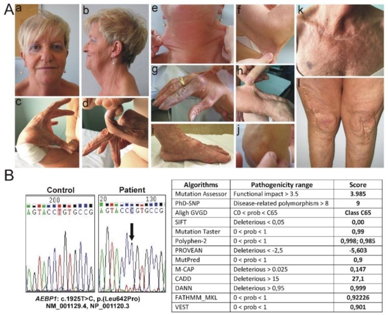
Ehlers-Danlos syndrome (EDS) comprises clinically heterogeneous connective tissue disorders with diverse molecular etiologies. The 2017 International Classification for EDS recognized 13 distinct subtypes caused by pathogenic variants in 19 genes mainly encoding fibrillar collagens and collagen-modifying or processing proteins. Recently, a new EDS subtype, i.e., classical-like EDS type 2, was defined after the identification, in six patients with clinical findings reminiscent of EDS, of recessive alterations in AEBP1, which encodes the aortic carboxypeptidase⁻like protein associating with collagens in the extracellular matrix. Herein, we report on a 53-year-old patient, born from healthy second-cousins, who fitted the diagnostic criteria for classical EDS (cEDS) for the presence of hyperextensible skin with multiple atrophic scars, generalized joint hypermobility, and other minor criteria. Molecular analyses of cEDS genes did not identify any causal variant. Therefore, AEBP1 sequencing was performed that revealed homozygosity for the rare c.1925T>C p.(Leu642Pro) variant classified as likely pathogenetic (class 4) according to the American College of Medical Genetics and Genomics (ACMG) guidelines. The comparison of the patient's features with those of the other patients reported up to now and the identification of the first missense variant likely associated with the condition offer future perspectives for EDS nosology and research in this field.
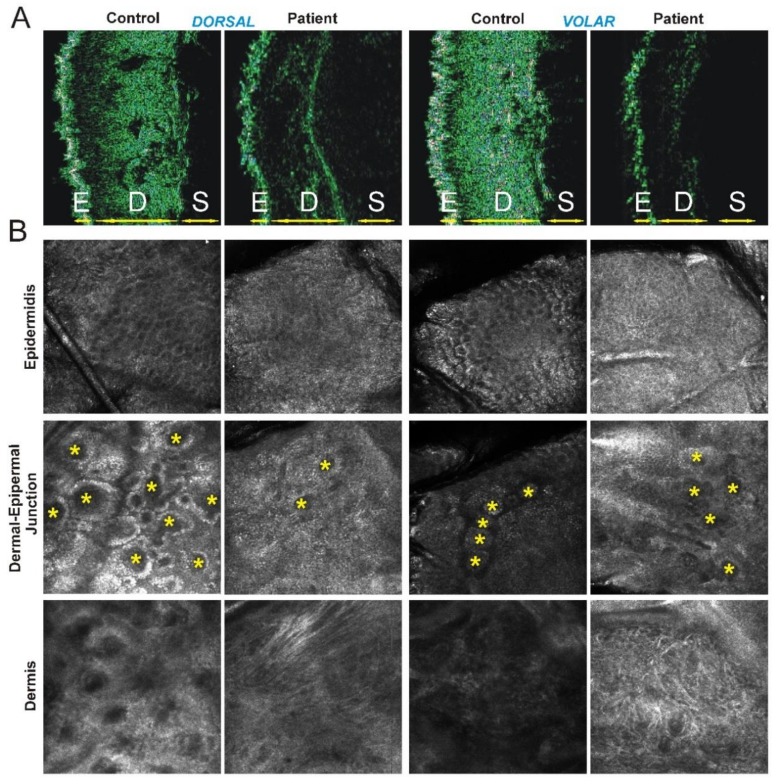
Keywords: AEBP1; aortic carboxypeptidase-like protein; classical Ehlers-Danlos syndrome; classical-like Ehlers-Danlos syndrome type 2; differential diagnosis; high-frequency ultrasonography; reflectance confocal microscopy.
Conflict of interest statement
All authors declare that there is no conflict of interest concerning this work.
Figures
Similar articles
-
Identification of the novel COL5A1 c.3369_3431dup, p.(Glu1124_Gly1144dup) variant in a patient with incomplete classical Ehlers-Danlos syndrome: The importance of phenotype-guided genetic testing.Mol Genet Genomic Med. 2020 Oct;8(10):e1422. doi: 10.1002/mgg3.1422. Epub 2020 Jul 28. Mol Genet Genomic Med. 2020. PMID: 32720758 Free PMC article.
-
Clinical and Molecular Characterization of a Novel Homozygous Frameshift Variant in AEBP1-Related Classical-like Ehlers Danlos Syndrome Type 2 with Comparison to Previously Reported Rare Cases.Genes (Basel). 2024 Apr 6;15(4):461. doi: 10.3390/genes15040461. Genes (Basel). 2024. PMID: 38674395 Free PMC article.
-
Bi-allelic AEBP1 mutations in two patients with Ehlers-Danlos syndrome.Hum Mol Genet. 2019 Jun 1;28(11):1853-1864. doi: 10.1093/hmg/ddz024. Hum Mol Genet. 2019. PMID: 30668708
-
The Ehlers-Danlos syndromes, rare types.Am J Med Genet C Semin Med Genet. 2017 Mar;175(1):70-115. doi: 10.1002/ajmg.c.31550. Am J Med Genet C Semin Med Genet. 2017. PMID: 28306225 Review.
-
Vascular aspects of the Ehlers-Danlos Syndromes.Matrix Biol. 2018 Oct;71-72:380-395. doi: 10.1016/j.matbio.2018.04.013. Epub 2018 Apr 27. Matrix Biol. 2018. PMID: 29709596 Review.
Cited by
-
Further Defining the Phenotypic Spectrum of B3GAT3 Mutations and Literature Review on Linkeropathy Syndromes.Genes (Basel). 2019 Aug 21;10(9):631. doi: 10.3390/genes10090631. Genes (Basel). 2019. PMID: 31438591 Free PMC article. Review.
-
An exemplary model of genetic counselling for highly specialised services.J Community Genet. 2023 Apr;14(2):115-119. doi: 10.1007/s12687-023-00640-4. Epub 2023 Mar 9. J Community Genet. 2023. PMID: 36892793 Free PMC article. Review.
-
Identification of the novel COL5A1 c.3369_3431dup, p.(Glu1124_Gly1144dup) variant in a patient with incomplete classical Ehlers-Danlos syndrome: The importance of phenotype-guided genetic testing.Mol Genet Genomic Med. 2020 Oct;8(10):e1422. doi: 10.1002/mgg3.1422. Epub 2020 Jul 28. Mol Genet Genomic Med. 2020. PMID: 32720758 Free PMC article.
-
Molecular Genetics and Pathogenesis of Ehlers-Danlos Syndrome and Related Connective Tissue Disorders.Genes (Basel). 2020 May 13;11(5):547. doi: 10.3390/genes11050547. Genes (Basel). 2020. PMID: 32414079 Free PMC article.
-
Clinical and Molecular Characterization of a Novel Homozygous Frameshift Variant in AEBP1-Related Classical-like Ehlers Danlos Syndrome Type 2 with Comparison to Previously Reported Rare Cases.Genes (Basel). 2024 Apr 6;15(4):461. doi: 10.3390/genes15040461. Genes (Basel). 2024. PMID: 38674395 Free PMC article.
References
-
- Beighton P., De Paepe A., Steinmann B., Tsipouras P., Wenstrup R.J. Ehlers-Danlos syndromes: Revised nosology, Villefranche, 1997. Ehlers-Danlos National Foundation (USA) and Ehlers-Danlos Support Group (UK) Am. J. Med. Genet. 1998;77:31–37. doi: 10.1002/(SICI)1096-8628(19980428)77:1<31::AID-AJMG8>3.0.CO;2-O. - DOI - PubMed
-
- Ritelli M., Dordoni C., Venturini M., Chiarelli N., Quinzani S., Traversa M., Zoppi N., Vascellaro A., Wischmeijer A., Manfredini E., et al. Clinical and molecular characterization of 40 patients with classic Ehlers-Danlos syndrome: Identification of 18 COL5A1 and 2 COL5A2 novel mutations. Orphanet J. Rare Dis. 2013;12:8–58. doi: 10.1186/1750-1172-8-58. - DOI - PMC - PubMed
-
- Colombi M., Dordoni C., Venturini M., Ciaccio C., Morlino S., Chiarelli N., Zanca A., Calzavara-Pinton P., Zoppi N., Castori M., et al. Spectrum of mucocutaneous, ocular and facial features and delineation of novel presentations in 62 classical Ehlers-Danlos syndrome patients. Clin. Genet. 2017;92:624–631. doi: 10.1111/cge.13052. - DOI - PubMed
Publication types
MeSH terms
Substances
Supplementary concepts
LinkOut - more resources
Full Text Sources
Medical
Miscellaneous
