Review
. 2019 Feb 12;8(2):154.
doi: 10.3390/cells8020154.

Role of Exosomes in the Regulation of T-cell Mediated Immune Responses and in Autoimmune Disease

Affiliations
Review

Role of Exosomes in the Regulation of T-cell Mediated Immune Responses and in Autoimmune Disease

Alberto Anel et al. Cells. .

Abstract

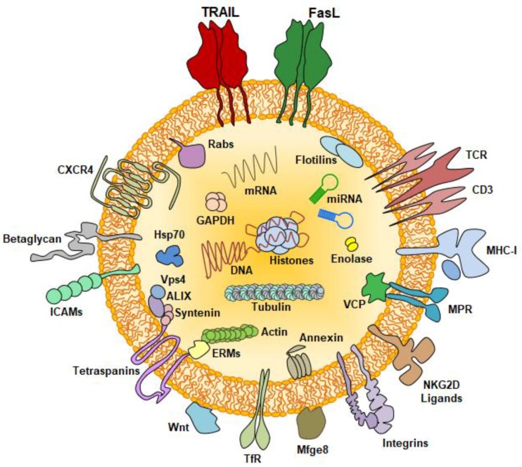
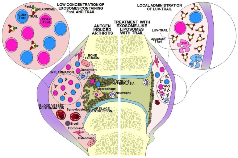
: T-cell mediated immune responses should be regulated to avoid the development of autoimmune or chronic inflammatory diseases. Several mechanisms have been described to regulate this process, namely death of overactivated T cells by cytokine deprivation, suppression by T regulatory cells (Treg), induction of expression of immune checkpoint molecules such as CTLA-4 and PD-1, or activation-induced cell death (AICD). In addition, activated T cells release membrane microvesicles called exosomes during these regulatory processes. In this review, we revise the role of exosome secretion in the different pathways of immune regulation described to date and its importance in the prevention or development of autoimmune disease. The expression of membrane-bound death ligands on the surface of exosomes during AICD or the more recently described transfer of miRNA or even DNA inside T-cell exosomes is a molecular mechanism that will be analyzed.

Keywords: autoimmunity; exosomes; extracellular vesicles; immune regulation.


Conflict of interest statement

The authors declare no conflict of interest.

Figures

Figure 1
Figure 1
Schematic representation of a T cell derived exosome, showing the location of several of the most important functional proteins expressed, together with miRNA and DNA.
Figure 2
Figure 2
Left, schematic description of the situation in an arthritic lesion with the prototypical pathologic hallmarks: inflammatory infiltrate, blood vessel neo-formation, synovial hyperplasia, and, as a consequence, bone erosion and cartilage destruction. This situation is associated with a low concentration of regulatory FasL and/or TRAIL-containing exosomes, probably favoring T cell chronic infiltration. Right, situation upon intra-articular delivery of large unilamellar vesicles decorated with recombinant TRAIL (LUV-TRAIL), resulting in the elimination of synovial hyperplasia and in a reduction of the inflammatory infiltrate (based on data in references [103,105]).

Similar articles

Cited by

References

    1. Kisielow P., Bluthmann H., Staerz U.D., Steinmetz M., von Boehmer H. Tolerance in T-cell-receptor transgenic mice involves deletion of nonmature CD4+8+ thymocytes. Nature. 1988;333:742–746. doi: 10.1038/333742a0. - DOI - PubMed
    1. Lenardo M., Chan F.K.M., Hornung F., MacFarland H., Siegel R., Wang J., Zheng L. Mature T lymphocyte apoptosis—Immune regulation in a dynamic and unpredictable antigenic enviroment. Annu. Rev. Immunol. 1999;17:221–253. doi: 10.1146/annurev.immunol.17.1.221. - DOI - PubMed
    1. Matzinger P. Tolerance, danger, and the extended family. Ann. Rev. Immunol. 1994;12:991–1045. doi: 10.1146/annurev.iy.12.040194.005015. - DOI - PubMed
    1. Schwartz R.H. A cell culture model for T lymphocyte clonal anergy. Science. 1990;248:1349–1356. doi: 10.1126/science.2113314. - DOI - PubMed
    1. Steinman R.M., Turley S., Mellman I., Inaba K. The induction of tolerance by dendritic cells that have captured apoptotic cells. J. Exp. Med. 2000;191:411–416. doi: 10.1084/jem.191.3.411. - DOI - PMC - PubMed

Publication types