Review
. 2019 Sep;25(3):245-263.
doi: 10.3350/cmh.2018.0090. Epub 2019 Feb 14.

Comparison of international guidelines for noninvasive diagnosis of hepatocellular carcinoma: 2018 update

Affiliations
Review

Comparison of international guidelines for noninvasive diagnosis of hepatocellular carcinoma: 2018 update

Tae-Hyung Kim et al. Clin Mol Hepatol. 2019 Sep.

Abstract

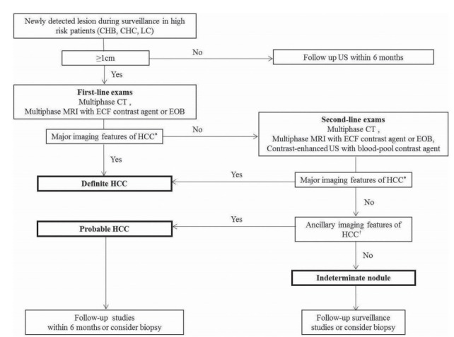
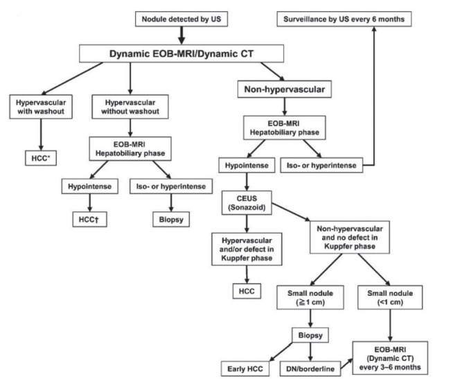
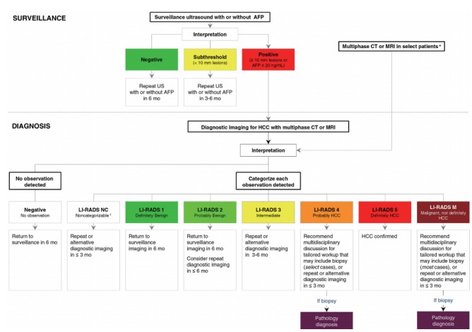
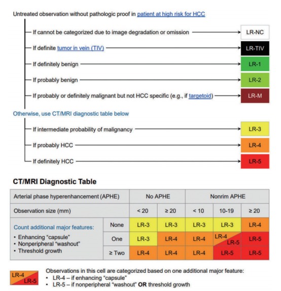
The goal of this review is to present the similarities and differences among the latest guidelines for noninvasive diagnosis of hepatocellular carcinoma (HCC) of American Association for the Study of Liver Disease (AASLD), European Association for the Study of the Liver (EASL), Liver Imaging Reporting and Data System (LI-RADS), Asian Pacific Association for the Study of the Liver (APASL), and Korean Liver Cancer Association- National Cancer Center (KLCA-NCC) of Korea. In 2018, major guideline updates have been proposed by the AASLD, EASL and KLCA-NCC; AASLD newly incorporated LI-RADS into their HCC diagnostic algorithm. The AASLD and EASL guidelines now include magnetic resonance imaging (MRI) using hepatobiliary contrast media as a first-line diagnostic test in addition to dynamic computed tomography and MRI using extracellular contrast media and the KLCA-NCC and EASL guidelines also include contrast-enhanced ultrasound as a second-line diagnostic test. We will comprehensively review the HCC surveillance and diagnostic algorithms and compare and highlight key features for each guideline. We also address limitations of current systems for the noninvasive diagnosis of HCC.

Keywords: Diagnosis; Guideline; Hepatocellular carcinoma; Standards.


Conflict of interest statement

Conflicts of Interest

Authors declare no conflicts of interest.

Figures

Figure 1.
Figure 1.
Diagnostic algorithm for a suspected hepatocellular carcinoma (HCC) using the new Korean Liver Cancer Association-National Cancer Center Korea practice guidelines. Adapted from KLCA-NCC guidelines [13]. CHB, chronic hepatitis B virus; CHC, chronic hepatitis C virus; LC, liver cirrhosis; ECF, extracellular fluid. *Major imaging features of HCC include arterial hyperenhancement and the wash-out appearance during portal venous, delayed, or hepatobiliary phases on multiphasic computed tomography (CT) or magnetic resonance imaging (MRI) using extracellular contrast agents or gadoxetate disodium (EOB) in nodules ≥1 cm in diameter. However, the lesion should not show either marked T2 high signal intensity or the targetoid appearance on diffusion weighted imaging (DWI) or contrast-enhanced sequences. On contrast-enhanced ultrasonography (US) as a second line exam, major imaging features include arterial hyperenhancement and late onset (≥ 60 seconds) mild wash-out; In nodule(s) with some but not all of the aforementioned major imaging features of HCC, the category of “probable” HCC can be assigned only when the lesion fulfills at least one item from each of the following two categories of ancillary imaging features. The two categories which make up ancillary imaging features are findings favoring malignancy in general (mild-to-moderate T2 hyperintensity, restricted diffusion, hepatobiliary phase hypointensity, interval growth) and those favoring HCC in particular (non-enhancing capsule, mosaic architecture, nodule-in-nodule appearance, fat or blood products in the mass). These criteria should be applied only to a lesion which shows neither marked T2 hyperintensity nor a targetoid appearance on diffusion-weighted images or contrast-enhanced sequences.
Figure 2.
Figure 2.
Diagnostic algorithm for hepatocellular carcinoma using multiple modalities according to Asian Pacific Association for the Study of the Liver (APASL). Reprint with permission from Omata et al [8]. US, ultrasonography; EOB, gadoxetate disodium; CT, computed tomography; HCC, hepatocellular carcinoma; CEUS, contrast enhanced ultrasonography; DN, dysplastic nodule. *Cavernous hemangioma sometimes shows hypointensity on the equilibrium (transitional) phase of dynamic Gd-EOB DTPA magnetic resonance imaging (MRI) (pseudo-wash-out). It should be excluded by further MRI sequences and/or other imaging modalities; Cavernous hemangioma usually shows hypointensity on the hepatobiliary phase of Gd-EOB DTPA MRI. It should be excluded by other MRI sequences and/or other imaging modalities.
Figure 3.
Figure 3.
Diagnostic algorithm for surveillance and diagnosis of hepatocellular carcinoma (HCC) according to American Association for the Study of Liver Disease (AASLD). Reprint with permission from Marrero et al.11 AFP, alphafeto protein; US, ultrasonography; mo, months; CT, computed tomography; MRI, magnetic resonance imaging; LI-RADS, Liver Imaging Reporting and Data System; NC, noncategorizable; M, malignancy (not necessarily HCC). *Some high-risk patients may undergo multiphase CT or MRI for HCC surveillance (depending on patient body habitus, visibility of liver at ultrasound, being on the transplant waiting list and other factors); These are due to technical problem such as image omission or severe degradation.
Figure 4.
Figure 4.
The computed tomography (CT)/magnetic resonance imaging (MRI) diagnostic algorithm of hepatocellular carcinoma (HCC) according to Liver Imaging Reporting and Data System (LI-RADS) v 2018. Reprint with permission from American College of Radiology.10 LR, lirads; NC, noncategorizable; TIV, tumor in vein; M, malignancy (not necessarily HCC).
Figure 5.
Figure 5.
Diagnostic algorithm and recall policy in cirrhotic liver according to European Association for the Study of the Liver (EASL). Reprint with permission from EASL.12 US, ultrasonography; mo, months; CT, computed tomography; MRI, magnetic resonance imaging; HCC, hepatocellular carcinoma. *Using extracellular magnetic resonance contrast agents or gadobenate dimeglumine; Using the following diagnostic criteria: arterial phase hyperenhancement (APHE) and washout on the portal venous phase; Using the following diagnostic criteria: APHE and mild washout after 60 seconds; §Lesion <1 cm stable for 12 months (three controls after four months) can be shifted back to regular 6 months surveillance; ||Optional for centre-based programmes.

Similar articles

Cited by

References

    1. Choo SP, Tan WL, Goh BKP, Tai WM, Zhu AX. Comparison of hepatocellular carcinoma in Eastern versus Western populations. Cancer. 2016;122:3430–3446. - PubMed
    1. Global Burden of Disease Liver Cancer Collaboration. Akinyemiju T, Abera S, Ahmed M, Alam N, Alemayohu MA, et al. The burden of primary liver cancer and underlying etiologies from 1990 to 2015 at the global, regional, and national level: results from the global burden of disease study 2015. JAMA Oncol. 2017;3:1683–1691. - PMC - PubMed
    1. Shields A, Reddy KR. Hepatocellular carcinoma: current treatment strategies. Curr Treat Options Gastroenterol. 2005;8:457–466. - PubMed
    1. Heimbach JK, Kulik LM, Finn RS, Sirlin CB, Abecassis MM, Roberts LR, et al. AASLD guidelines for the treatment of hepatocellular carcinoma. Hepatology. 2018;67:358–380. - PubMed
    1. Tang A, Cruite I, Mitchell DG, Sirlin CB. Hepatocellular carcinoma imaging systems: why they exist, how they have evolved, and how they differ. Abdom Radiol (NY) 2018;43:3–12. - PubMed