. 2019 Feb 13;19(1):147.
doi: 10.1186/s12885-019-5338-5.

Patient satisfaction of radiofrequency ablation for symptomatic benign solid thyroid nodules: our experience for 2-year follow up

Affiliations

Patient satisfaction of radiofrequency ablation for symptomatic benign solid thyroid nodules: our experience for 2-year follow up

Yang Guang et al. BMC Cancer. .

Abstract

Background: The patient satisfaction of symptoms improvement and disease factors that may affect long-term treatment efficacy of radiofrequency ablation (RFA) for non-functioning solid benign thyroid nodules (TNs) over a 2-year follow up study was investigated.

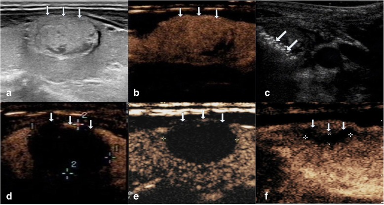
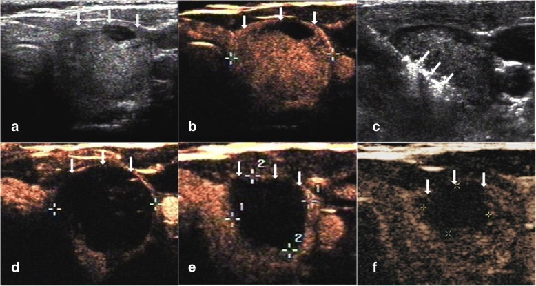
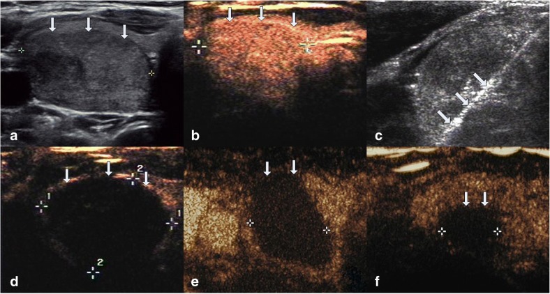
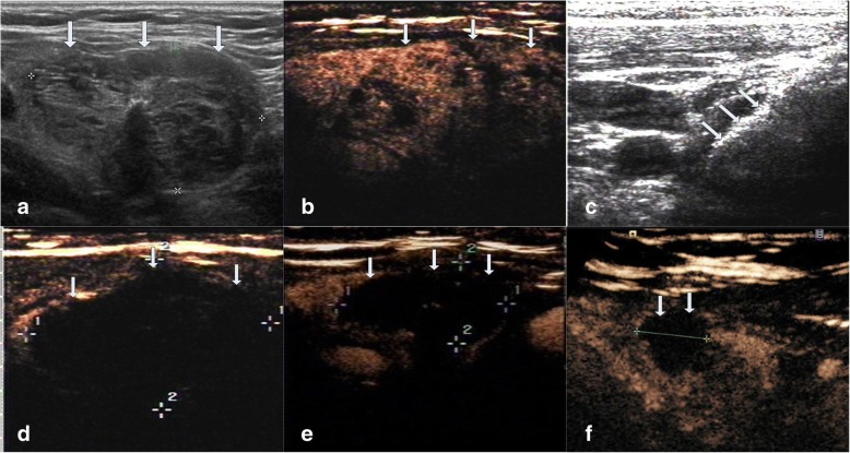
Methods: This retrospective study evaluated 194 non-functioning solid benign TNs of 103 patients. The TNs were categorized as small (≤5 ml), medium (5.1 to 13 ml), intermediate (13.1 to 30 ml) and large (over 30 ml) according to the initial volume of TNs before ablation. Clinical evaluation and contrast-enhanced ultrasound (CEUS) were carried out before ablation and the follow up at 1, 3, 6 months and every 6 months after ablation. All patients were asked to assess the cosmetic score (1-4 scores) and symptom score (0-10 scores) before ablation and every follow up after ablation.

Results: All patients underwent RFA without any major complications. The mean treatment sessions were 1.5 ± 0.6. 98 nodules required a single session (98/194, 50.5%), 87 required two sessions (87/194, 44.9%), 9 required three sessions (9/194, 4.6%). The average follow up months were 16.3 ± 5.6 (range, 6-24 months) and no nodule regrew in our study. After RFA treatment, the TNs volume significantly decreased (P < 0.001). The small group of nodules shrunk larger compared to the medium, intermediate and large groups (P < 0.001). Cosmetic signs and pressure symptoms were significantly improved, particularly in the intermediate and large groups (P < 0.05).

Conclusions: RFA is effective for treating non-functioning solid benign TNs and controlling clinical symptoms with a low complication rate during 2 years follow up. The reduction rate was related to the initial volume of nodules. Patients were satisfied with cosmetic signs and pressure symptoms improvement, particularly in the intermediate and large groups. However, multiple RFA treatments should be used in larger nodules to achieve the desired clinical outcomes.

Keywords: Radiofrequency ablation (RFA); Thyroid nodules; Ultrasound.


Conflict of interest statement

Ethics approval and consent to participate

The protocol was approved by the Institutional Review Board of the Beijing Tian Tan Hospital Ethics Committee and the Chinese PLA General Hospital Ethics Committee. All subjects provided written informed consent.

Consent for publication

Not applicable.

Competing interests

The authors declare that they have no competing interests.

Publisher’s Note

Springer Nature remains neutral with regard to jurisdictional claims in published maps and institutional affiliations.

Figures

Fig. 1
Fig. 1
Changes in VRR of the four groups of 194 non-functioning solid benign thyroid nodules at each follow-up
Fig. 2
Fig. 2
Images of RFA treatment and follow-up of a thyroid nodule at the left lobe in small group. a Conventional ultrasound image shows a solid thyroid nodule. b CEUS performed before RFA shows an irregular enhancement in nodule (white arrows). c During RFA, ultrasound monitoring of the procedure shows gas formation in the nodule which covered by a hyperechoic ablation area (white arrows). d CEUS performed immediately after RFA shows a complete lack of enhancement in the treated area (white arrows). e One month after RFA, CEUS shows the treated area shrunk and that complete lack of enhancement (white arrows). f 1-year after RFA, CEUS shows complete lack of enhancement in the treated area, remained as a small scarlike lesion (white arrows)
Fig. 3
Fig. 3
Images of RFA treatment and follow-up of a thyroid nodule at left lobe in medium group. a Conventional ultrasound image shows a thyroid nodule with solid portion over 80%. b CEUS performed before RFA shows an irregular enhancement in nodule with a regular hyperechoic ring around the nodule (white arrows). c During RFA, ultrasound monitoring of the procedure shows gas formation in the nodule which covered by a hyperechoic ablation area (white arrows). d CEUS performed immediately after RFA shows a complete lack of enhancement in the treated area (white arrows). e One month after RFA, CEUS shows the treated area shrunk and that complete lack of enhancement (white arrows). f 1-year after RFA, CEUS shows complete lack of enhancement in the treated area, remained as a small scarlike lesion (white arrows)
Fig. 4
Fig. 4
Images of RFA treatment and follow-up of a thyroid nodule at right lobe in intermediate group. a Conventional ultrasound image shows a solid thyroid nodule. b CEUS performed before RFA shows an irregular enhancement in nodule (white arrows). c During RFA, ultrasound monitoring of the procedure shows gas formation in the nodule which covered by a hyperechoic ablation area (white arrows). d CEUS performed immediately after RFA shows a complete lack of enhancement in the treated area (white arrows). e Three months after RFA, CEUS shows the treated area shrunk and that complete lack of enhancement (white arrows). f 1.5-year after RFA, CEUS shows complete lack of enhancement in the treated area, remained as a small scarlike lesion (white arrows)
Fig. 5
Fig. 5
Images of RFA treatment and follow-up of a thyroid nodule at right lobe in large group. a Conventional ultrasound image shows a thyroid nodule with solid portion over 80%. b CEUS performed before RFA shows an irregular enhancement in nodule (white arrows). c During RFA, ultrasound monitoring of the procedure shows gas formation in the nodule which covered by a hyperechoic ablation area (white arrows). d CEUS performed immediately after RFA shows a complete lack of enhancement in the treated area (white arrows). e Six months after RFA, CEUS shows the treated area shrunk and that complete lack of enhancement (white arrows). f 2-year after RFA, CEUS shows complete lack of enhancement in the treated area, remained as a small scarlike lesion (white arrows)

References

    1. Guth S, Theune U, Aberle J, Galach A, Bamberger CM. Very high prevalence of thyroid nodules detected by high frequency (13 MHz) ultrasound examination. Eur J Clin Investig. 2009;39:699–706. doi: 10.1111/j.1365-2362.2009.02162.x. - DOI - PubMed
    1. Smith-Bindman R, Miglioretti DL, Johnson E, Lee C, Feigelson HS, Flynn M, et al. Use of diagnostic imaging studies and associated radiation exposure for patients enrolled in large integrated health care systems, 1996-2010. JAMA. 2012;307:2400–2409. doi: 10.1001/jama.2012.5960. - DOI - PMC - PubMed
    1. Alexander EK, Hurwitz S, Heering JP, Benson CB, Frates MC, Doubilet PM, et al. Natural history of benign solid and cystic thyroid nodules. Ann Intern Med. 2003;138:315–318. doi: 10.7326/0003-4819-138-4-200302180-00010. - DOI - PubMed
    1. Kihara M, Hirokawa M, Masuoka H, Yabuta T, Shindo H, Higashiyama T, et al. Evaluation of cytologically benign solitary thyroid nodules by ultrasonography: a retrospective analysis of 1877 cases. Auris Nasus Larynx. 2013;40:308–311. doi: 10.1016/j.anl.2012.09.007. - DOI - PubMed
    1. Lee S, Skelton TS, Zheng F, Schwartz KA, Perrier ND, Lee JE, et al. The biopsy-proven benign thyroid nodule: is long-term follow-up necessary? J Am Coll Surg. 2013;217:81–88. doi: 10.1016/j.jamcollsurg.2013.03.014. - DOI - PubMed

LinkOut - more resources