. 2019 Feb 13;7(1):45.
doi: 10.1186/s40425-019-0521-4.

The IL-1/IL-1 receptor axis and tumor cell released inflammasome adaptor ASC are key regulators of TSLP secretion by cancer associated fibroblasts in pancreatic cancer

Affiliations

The IL-1/IL-1 receptor axis and tumor cell released inflammasome adaptor ASC are key regulators of TSLP secretion by cancer associated fibroblasts in pancreatic cancer

Emanuela Brunetto et al. J Immunother Cancer. .

Abstract

Background: The thymic stromal lymphopoietin (TSLP), a key cytokine for development of Th2 immunity, is produced by cancer associated fibroblasts (CAFs) in pancreatic cancer where predominant tumor infiltrating Th2 over Th1 cells correlates with reduced patients' survival. Which cells and molecules are mostly relevant in driving TSLP secretion by CAFs in pancreatic cancer is not defined.

Methods: We performed in vitro, in vivo and ex-vivo analyses. For in vitro studies we used pancreatic cancer cell lines, primary CAFs cultures, and THP1 cells. TSLP secretion by CAFs was used as a read-out system to identify in vitro relevant tumor-derived inflammatory cytokines and molecules. For in vivo studies human pancreatic cancer cells and CAFs were orthotopically injected in immunodeficient mice. For ex-vivo studies immunohistochemistry was performed to detect ASC (apoptosis-associated speck-like protein containing a caspase recruitment domain) expression in surgical samples. Bioinformatics was applied to interrogate published data sets.

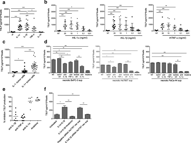
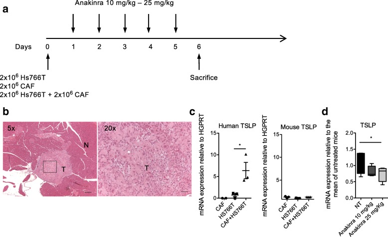
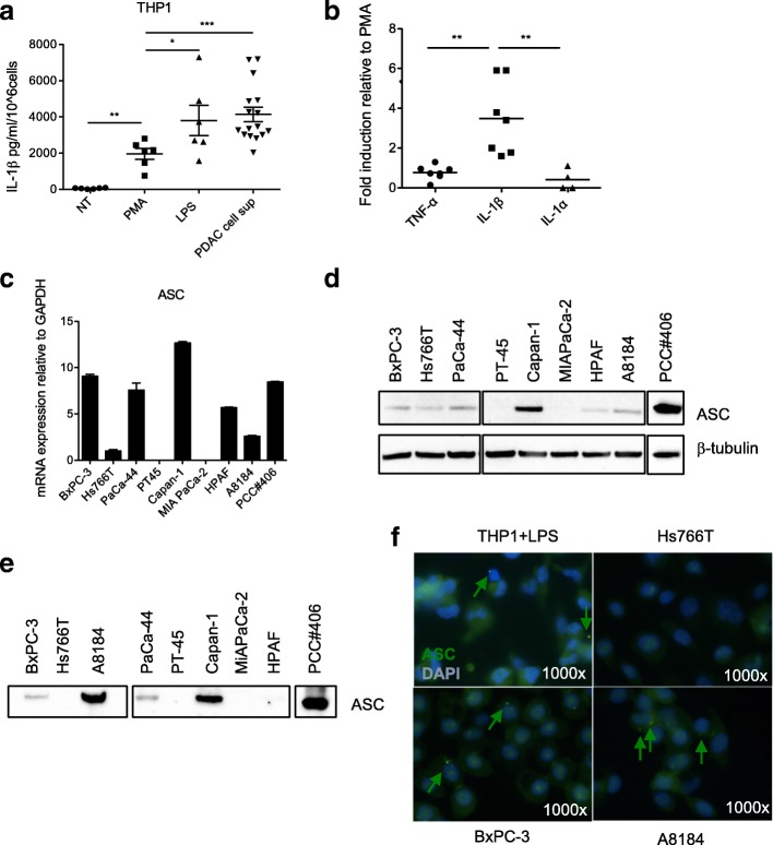
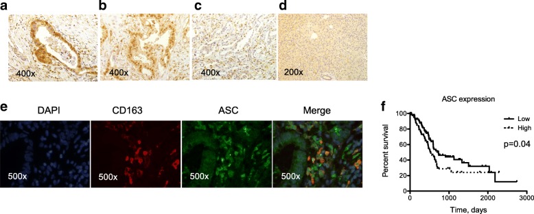
Results: We show in vitro that IL-1α and IL-1β released by pancreatic cancer cells and tumor cell-conditioned macrophages are crucial for TSLP secretion by CAFs. Treatment of immunodeficient mice orthotopically injected with human IL-1 positive pancreatic cancer cells plus CAFs using the IL-1R antagonist anakinra significantly reduced TSLP expression in the tumor. Importantly, we found that pancreatic cancer cells release alarmins, among which ASC, able to induce IL-1β secretion in macrophages. The relevance of ASC was confirmed ex-vivo by its expression in both tumor cells and tumor associated macrophages in pancreatic cancer surgical samples and survival data analyses showing statistically significant inverse correlation between ASC expression and survival in pancreatic cancer patients.

Conclusions: Our findings indicate that tumor released IL-1α and IL-1β and ASC are key regulators of TSLP secretion by CAFs and their targeting should ultimately dampen Th2 inflammation and improve overall survival in pancreatic cancer.

Keywords: Cancer associated fibroblasts; IL-1; Inflammasome; Inflammasome adaptor ASC (apoptosis-associated speck-like protein containing a caspase-activating recruitment domain); Pancreatic cancer; Th2 inflammation; Thymic stromal lymphopoietin.


Conflict of interest statement

Ethics approval and consent to participate

The Institutional Ethics Committee (Comitato Etico Fondazione Centro San Raffaele, Istituto Scientifico Ospedale San Raffaele) had approved the study protocol and written informed consent was obtained from all patients.

Consent for publication

Not applicable

Competing interests

The authors declare that they have no competing interests.

Publisher’s Note

Springer Nature remains neutral with regard to jurisdictional claims in published maps and institutional affiliations.

Figures

Fig. 1
Fig. 1
Tumor cell-derived IL-1 has a key role in TSLP secretion by CAFs. a TSLP secretion by CAFs (n = 18) either untreated (NT) or treated with the indicated cytokines (20 ng/ml). Each symbol refers to a single experiment and represents the mean of duplicate determinations. Significance was determined using Mann Whitney test. b TSLP secretion by CAFs in the absence (NT) or in the presence of increasing doses of recombinant IL-1α, IL-1β and TNF-α. Significance was determined using Mann Whitney test. c TSLP secretion by CAFs (n = 14) either untreated (NT) or treated with supernatants of necrotic IL-1 negative (IL-1-) (n = 16) or positive (IL-1+) (n = 26) tumor cells. Each symbol refers to a single experiment and represents the mean of duplicate determinations. Significance was determined using Mann Whitney test. d TSLP secretion by CAFs induced by necrotic tumor cell-derived supernatants in the absence (NT) or in the presence of the indicated neutralizing Abs or isotype control Abs or the IL-1RA (Anakinra). Representative experiments performed with the indicated tumor cell lines. Error bars represent standard deviations of triplicate values. Significance was determined using Student’s t-test. e Cumulative results: each dot refers to a single experiment and represents the percentage of inhibition calculated on the value of TSLP secretion in the presence of the isotype control antibody. f TSLP secretion by CAFs either untreated or stimulated with the supernatant of necrotic Hs766T cells after treatment with IL-1α/IL-1β siRNA or negative control siRNA. Treatment of CAFs with recombinant IL-1α + IL-1β (20 ng/ml each) in the absence or in the presence of anakinra were used as controls. Error bars represent standard deviations of triplicate values. Significance was determined using Student’s t-test. Values significantly different were indicated as: *p < 0.05, **p < 0.01 and ***p < 0.001
Fig. 2
Fig. 2
Treatment of immunodeficient NSG mice orthotopically transplanted with IL-1 positive Hs766T cells plus CAFs with anakinra reduces TSLP expression in vivo. a Treatment schedule. Mice were orthotopically injected on day 0 with Hs766T cells plus CAFs. Hs766T cells alone and CAFs alone were injected as controls. The next day mice transplanted with Hs766T cells plus CAFs started intraperitoneal injection with the two indicated doses of anakinra and mice were sacrificed on day 6 after daily administration. b Hematoxylin and eosin staining of the pancreas. Left, a tumor mass (T) localized within the normal pancreas (N) is evident. Right, at higher magnification, histological features of pancreatic cancer with cancer cells, fibroblasts and extracellular matrix can be recognized. c Human (left) and mouse (right) TSLP mRNA expression in pancreata of NSG mice implanted with CAFs alone (n = 3), Hs766T cells alone (n = 3) or CAFs plus Hs766T cells (n = 3). Significance was determined using Student’s t-test. d TSLP expression in pancreata of NSG mice untreated (NT) (n = 4) or treated with the two indicated doses of Anakinra (n = 4 for each dose). Significance was determined using Student’s t-test. Values significantly different were indicated as: *p < 0.05
Fig. 3
Fig. 3
Tumor cells induce IL-1β secretion in macrophages through the release of alarmins, among which ASC. a IL-1β secretion by THP-1 cells after priming with PMA and stimulation with tumor cell-derived supernatants (PDAC cell sup) (n = 16). LPS was used as positive control. Each symbol refers to a single experiment and represents the mean of duplicate determinations. Significance was determined using Mann Whitney test. b Comparison of cytokine secretion by THP1 treated with PDAC cell sup, as in a. Each symbol refers to a single experiment and represent the mean of duplicate determinations. The values are reported as fold increase relative to those of THP1 stimulated in the presence of PMA only. Significance was determined using Mann Whitney test. c ASC mRNA expression in the indicated PDAC cell lines. d WB analysis of ASC protein in the indicated PDAC cell lines. e WB analysis to determine levels of ASC protein in TCA precipitated supernatants of the indicated PDAC cell lines. f Immunofluorescence analysis of ASC expression in LPS treated THP-1 (positive control) and the indicated PDAC cell lines. ASC aggregates/specks are represented by green dots (arrows)
Fig. 4
Fig. 4
Tumor cell-derived ASC induces IL-1β secretion by macrophages and indirectly TSLP secretion by CAFs. a IL-1β secretion by THP-1 cells in the presence of the indicated stimuli, i.e. none (NT), PMA alone, PMA + LPS, PMA+ tumor cell sup (untreated sup), ASC-depleted or iso-rab-depleted tumor cell sups. The experiments were performed with BxPC3 (left) and A8184 (right) cell lines. Error bars represent standard deviations of triplicate values. Significance was determined using Students’ t test. b Cumulative results of independent experiments (each symbol refers to a single experiment). Percentage of inhibition was calculated on the values of THP1 in the presence of untreated sup. Significance was determined using paired Students’ t test. c IL-1β secretion by THP-1 cells in the presence of the indicated stimuli, i.e. none (NT), PMA alone, PMA + LPS, the supernatant (sup) of tumor cells (BxPC3 and A8184) either mock transfected (mock sup) or transfected with negative control siRNA (neg ctrl siRNA) or ASC siRNA. Error bars represent standard deviations of triplicate values. Significance was determined using Students’ t test. d Cumulative results of independent experiments (each symbol refers to a single experiment). Percentage of inhibition was calculated on the values of THP1 in the presence of mock-sup. Significance was determined using paired Students’ t test. e TSLP secretion by CAFs stimulated with the supernatant of THP1 cells treated with the supernatant of the indicated tumor cell lines (BxPC-3 and A8184) either mock transfected (mock sup) or transfected with control siRNA (neg ctrl sup) or ASC siRNA sup (n = 3). Error bars represent standard deviations of triplicate values. Significance was determined using unpaired Students’ t test. Values significantly different were indicated as: *p < 0.05, **p < 0.01 and ***p < 0.001
Fig. 5
Fig. 5
ASC is expressed in situ in tumor cells and TAMs and its expression in primary tumors correlates with reduced overall survival. a-d Immunohistochemistry of ASC protein expression in representative samples of tumor and tumor surrounding tissue. a ASC staining in both the nucleus and the cytoplasm in both tumor cells and TAMs. b ASC staining in both the nucleus and the cytoplasm in tumor cells while prevalently cytoplasmic in TAMs. c ASC staining is negative in tumor cells and in TAMs is prevalently nuclear. d ASC staining in normal pancreatic tissue surrounding the tumor. e Immunofluorescence staining of ASC (green) in tumor cells and TAMs (identified by CD163 staining in red). Cells co-expressing ASC and CD163 are indicated in orange (merge). f Kaplan-Meier curves of overall survival of 178 PDAC patients grouped based on median ASC RNA expression. Significance was evaluated by the Log-rank (Mantel-Cox) test (p = 0.04)

References

    1. Hidalgo M. Pancreatic cancer. N Engl J Med. 2010;362(17):1605–1617. - PubMed
    1. Conroy T, Desseigne F, Ychou M, Bouche O, Guimbaud R, Becouarn Y, Adenis A, Raoul JL, Gourgou-Bourgade S, de la Fouchardiere C, et al. FOLFIRINOX versus gemcitabine for metastatic pancreatic cancer. N Engl J Med. 2011;364(19):1817–1825. - PubMed
    1. Von Hoff DD, Ervin T, Arena FP, Chiorean EG, Infante J, Moore M, Seay T, Tjulandin SA, Ma WW, Saleh MN, et al. Increased survival in pancreatic cancer with nab-paclitaxel plus gemcitabine. N Engl J Med. 2013;369(18):1691–1703. - PMC - PubMed
    1. Hanahan D, Weinberg RA. Hallmarks of cancer: the next generation. Cell. 2011;144(5):646–674. - PubMed
    1. Balkwill FR, Mantovani A. Cancer-related inflammation: common themes and therapeutic opportunities. Semin Cancer Biol. 2012;22(1):33–40. - PubMed

Publication types

MeSH terms