Review
. 2019 Feb 13;32(2):e00075-18.
doi: 10.1128/CMR.00075-18. Print 2019 Mar 20.

Echinococcosis: Advances in the 21st Century

Affiliations
Review

Echinococcosis: Advances in the 21st Century

Hao Wen et al. Clin Microbiol Rev. .

Abstract

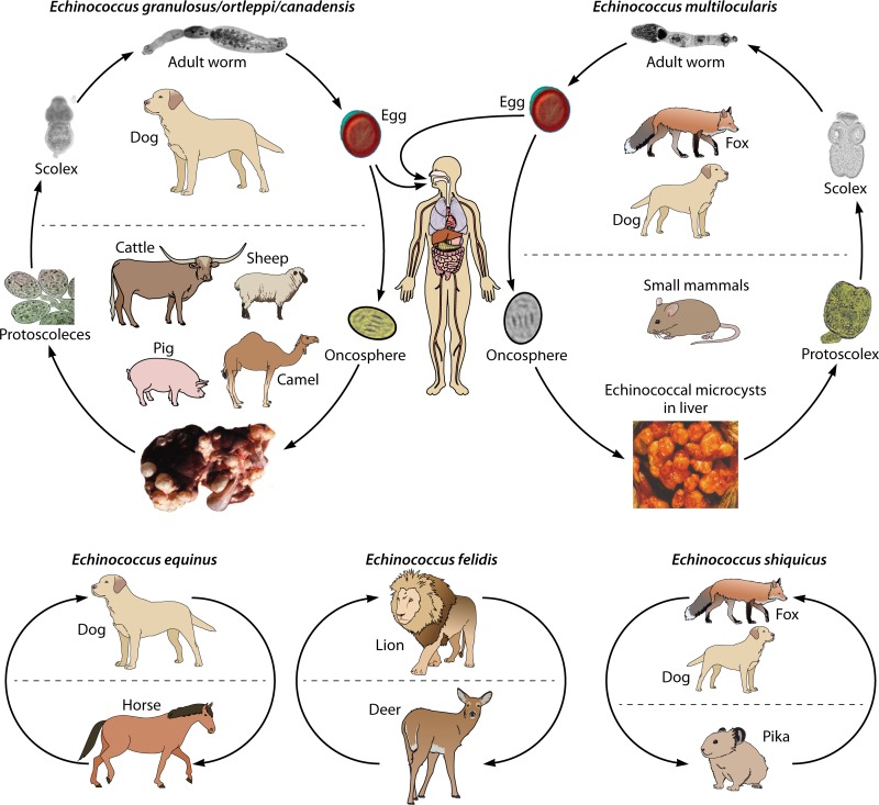
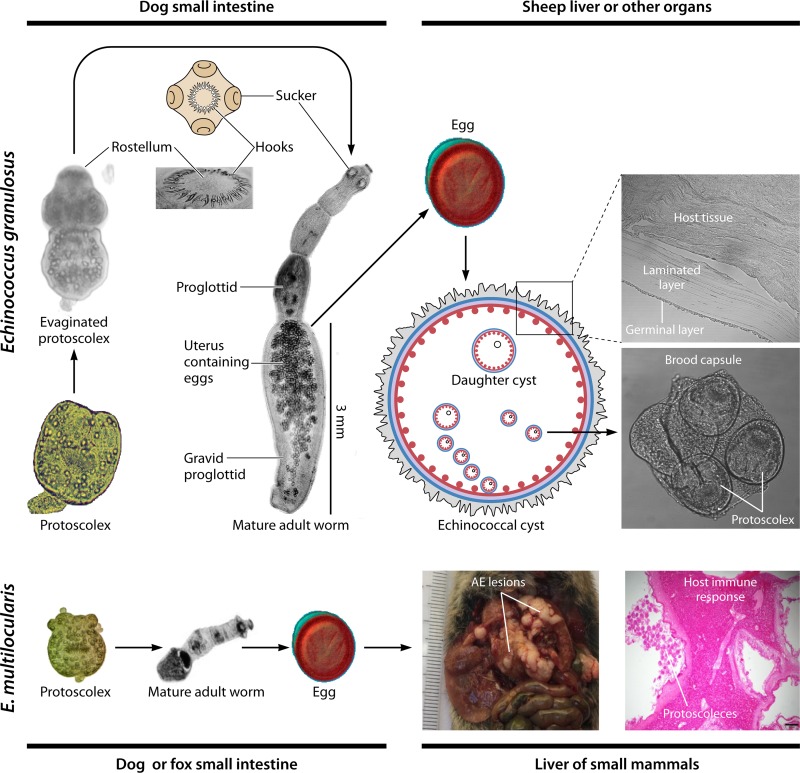
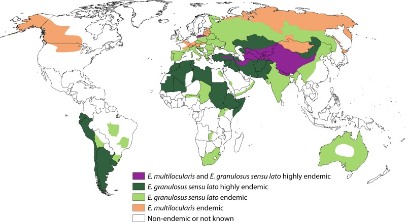
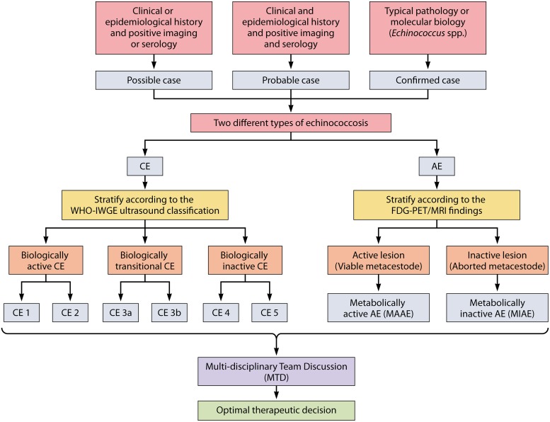
Echinococcosis is a zoonosis caused by cestodes of the genus Echinococcus (family Taeniidae). This serious and near-cosmopolitan disease continues to be a significant public health issue, with western China being the area of highest endemicity for both the cystic (CE) and alveolar (AE) forms of echinococcosis. Considerable advances have been made in the 21st century on the genetics, genomics, and molecular epidemiology of the causative parasites, on diagnostic tools, and on treatment techniques and control strategies, including the development and deployment of vaccines. In terms of surgery, new procedures have superseded traditional techniques, and total cystectomy in CE, ex vivo resection with autotransplantation in AE, and percutaneous and perendoscopic procedures in both diseases have improved treatment efficacy and the quality of life of patients. In this review, we summarize recent progress on the biology, epidemiology, diagnosis, management, control, and prevention of CE and AE. Currently there is no alternative drug to albendazole to treat echinococcosis, and new compounds are required urgently. Recently acquired genomic and proteomic information can provide a platform for improving diagnosis and for finding new drug and vaccine targets, with direct impact in the future on the control of echinococcosis, which continues to be a global challenge.

Keywords: Echinococcus; Echinococcus granulosus; Echinococcus multilocularis; alveolar echinococcosis; cystic echinococcosis; echinococcosis; genetic epidemiology; genome; strains/genotypes; transcriptome; zoonosis.


Figures

FIG 1
FIG 1
Life cycles of Echinococcus spp. Species responsible for human infection (E. granulosus sensu stricto, E. ortleppi, and E. canadensis [belonging to E. granulosus sensu lato] and E. multilocularis) are shown at the top. Species at the bottom (E. shiquicus, a species close to E. multilocularis, and E. equinus and E. felidis, belonging to E. granulosus sensu lato) are not known to cause disease in humans. Only the most common definitive and intermediate hosts which play a major role in life cycle/transmission are shown; other hosts may be encountered (especially wildlife hosts for E. granulosus sensu lato and domestic hosts for E. multilocularis). E. vogeli and E. oligarthra, which are responsible for polycystic echinococcosis in humans in Central and South America, are not represented in the figure.
FIG 2
FIG 2
Different developmental stages in Echinococcus granulosus and E. multilocularis. Growth of the larval cyst is unlimited, and it can, for E. granulosus, grow to 30 cm or more in humans, while the adult worm, egg, and protoscolex are limited in size and shape. Echinococcus sp. tapeworms have no gut, circulatory, or respiratory organs and have a highly adapted relationship with their mammalian hosts which they exploit for nutrients, signaling pathways, and neuroendocrine hormones. Strobilization is a notable feature of cestode biology, whereby proglottids (segments) bud distally from the anterior scolex, resulting in the production of tandem reproductive units (proglottids) exhibiting increasing degrees of development. Echinococcus is monoecious, and the last segment (gravid proglottid) produces diploid eggs that give rise to ovoid embryos, the oncospheres. However, a striking feature of the biology of Echinococcus is that the protoscolex has the potential to develop in either of two directions: it may develop into an adult tapeworm producing sexually produced eggs in the dog gut, or, if a hydatid cyst ruptures within the intermediate or human host, each released protoscolex is capable of differentiating asexually into a new cyst, a process termed “secondary” echinococcosis. While a unilocular fluid-filled bladder (cyst) is a feature of E. granulosus sensu lato in its larval stage, the metacestode of E. multilocularis consists of a mass of small, multilocular vesicles embedded in the immune reaction of the host (granuloma and fibrosis). These multiple and aggregated vesicles grow by proliferation of cells in the germinal layer of the metacestode.
FIG 3
FIG 3
Global distribution of Echinococcus granulosus sensu lato, responsible for cystic echinococcosis (CE), and Echinococcus multilocularis, responsible for alveolar echinococcosis (AE). The map is based on recent epidemiological studies (1, 13, 19, 247) as far as the current situation has been studied in a given area. The different colors represent a proxy for human prevalence and infection in animal hosts in a given area (to take autochthonous human cases into account only). For AE, the represented disease density is based mainly on the presence of autochthonous AE cases in humans, E. multilocularis metacestodes in small mammals, and E. multilocularis adult worms in foxes and dogs. For CE, the represented disease density is based mainly on the presence of autochthonous human cases of CE and of E. granulosus sensu lato metacestodes (irrespective of species or genotype) in intermediate hosts, including sheep, cattle, equids, and camels. For more accurate and detailed data and maps, see a recent comprehensive review paper by Deplazes et al. (13).
FIG 4
FIG 4
Algorithm for the diagnosis of cystic echinococcosis (CE) and alveolar echinococcosis (AE). Definitions of “possible,” “probable,” and “confirmed” cases refer to the “Expert consensus for diagnosis and treatment of echinococcosis in humans” (86). CE1 to CE5 refer to the “WHO-IWGE [World Health Organization Informal Working Group on Echinococcosis] international classification of ultrasound images in cystic echinococcosis for application in clinical and field epidemiological settings” (86) and Fig. 5. FDG-PET, fluorodeoxyglucose-positron emission tomography (increased uptake of FDG by the periparasitic immune response is the currently accepted evidence for AE lesion metabolic activity) (94). MRI, magnetic resonance imaging (identification of typical microcysts on T2-weighted images at MRI is a surrogate marker for AE lesion metabolic activity) (98).
FIG 5
FIG 5
Imaging of cystic echinococcosis. The description of ultrasound images is according to the WHO Informal Working Group on Echinococcosis (WHO-IWGE) international classification (86) and corresponding images were obtained from plain computed tomography (CT) scanning and T2-weighted magnetic resonance imaging (MRI) in representative cases. In the international classification, types CE1 and CE2 correspond to “active stages,” types CE3a and b to the “transitional stage,” and types CE4 and CE5 to “degenerating stages” (CT and MRI images provided by Liu Wenya, Department of Radiology, First Affiliated Hospital of Xinjiang Medical University, Urumqi, People’s Republic of China).
FIG 6
FIG 6
Complementarity of imaging techniques for the diagnosis and preoperative assessment of alveolar echinococcosis (AE) lesions in a patient with portal vein and bile duct invasion by the parasitic lesions. (A) On computed tomography (CT) scanning, the lesion shows the characteristic heterogeneous aspect of AE, with hyperdense calcifications (white arrow) and hypodense area (central necrosis) (black arrow). (B) Fat-suppressed T1-weighted magnetic resonance (MR) image after gadolinium injection at the portal venous phase shows an atrophy of the left liver and invasion of the left portal vein and of the left intrahepatic biliary tree. The lesion is at the contact of the gallbladder (white star) which is not invaded. (C) T2-weighted MR images show the presence of hyperintense microcysts (white arrows), pathognomonic of AE, but also a solid component (Kodama type II). (D) Fluorodeoxyglucose (FDG) uptake in positron emission tomography (PET) is markedly increased at the periphery of the lesion (white arrows). (E) Assessment of biliary tree involvement and treatment by perendoscopic stenting in a patient with late postoperative biliary complications of AE. Endoscopic retrograde cholangiopancreatography (ERCP) was performed (with a colonoscope because of previous gastrectomy), showing dilation of the extrahepatic and intrahepatic biliary tree with several defects (black arrows) because of biliary stones due to chronic biliary obstruction and bacterial superinfection. (F) Perendoscopic stenting via ERCP. After balloon dilation followed by extensive lavage with isotonic saline and stone extraction, 3 plastic stents (size, 7 and 10 French) are inserted in the stenosis of the bile duct (endoscopic view).
FIG 7
FIG 7
Algorithm for the treatment of cystic echinococcosis (CE), based on “WHO-IWGE international classification of ultrasound images in cystic echinococcosis for application in clinical and field epidemiological settings” (86) and Fig. 4 and 5. WHO-IWGE, World Health Organization Informal Working Group on Echinococcosis; ABZ, albendazole; PAIR, puncture-aspiration-injection-reaspiration (nonsurgical percutaneous interventional technique for treatment of CE cysts).
FIG 8
FIG 8
Schematic structure of the echinococcal cyst and different approaches for surgical removal. (A) The echinococcal cyst is made up of the adventitial layer, laminated layer, and germinal layer (from outside to inside). (B) Total cystectomy involves resection of the entire adventitial layer (“subadventitial resection”), the laminated layer, and the germinal layer. (C) Sub-total cystectomy involves partial resection of the adventitial layer and total resection of the laminated layer and the germinal layer, leaving parts of the adventitial layer in place whenever the operation is difficult because of the proximity of large vessels and/or adhesions. (D) Hepatectomy involves the en bloc resection of the echinococcal cyst along with part of the normal liver parenchyma. Partial cystectomy, which requires opening of the cyst, may leave all or part of the laminated layer and germinal layer and relies on the efficacy of a protoscolecide to destroy the metacestode; it should generally not be considered because of the potential for recurrence.
FIG 9
FIG 9
Algorithm for the treatment of alveolar echinococcosis. FDG-PET, fluorodeoxyglucose-positron emission tomography (increased uptake of FDG by the periparasitic immune response is the currently accepted evidence for AE lesion metabolic activity) (94). MRI, magnetic resonance imaging (identification of typical microcysts on T2-weighted images at MRI is a surrogate marker for AE lesion metabolic activity) (98). ABZ, albendazole; ELRA, ex vivo liver resection with autotransplantation.
FIG 10
FIG 10
Three-dimensional reconstruction in the application of ex vivo liver resection and autotransplantation (ELRA). (A) Portal veins, hepatic veins, and segments of liver are visualized by three-dimensional reconstruction. (B) Calculation of remnant hepatic parenchymal volume after three-dimensional reconstruction and virtual resection. The volume of remnant liver is 1,065.83 cm3 (yellow, giant AE lesion; blue, normal parenchyma). (C) Precise resection of giant AE lesion on bench (black arrow, normal liver parenchyma after resection). (D) The length (68.1 mm) of obliterated retro-hepatic vena cava is calculated through three-dimensional reconstruction (yellow, giant AE lesion; blue, retro-hepatic vena cava). (E) Hepaticojejunostomy is performed on the table (black arrow, anastomosis). (F) Postoperative follow-up demonstrating liver remnant and vasculatures (1, aorta and hepatic artery; 2, liver; 3, portal vein; 4, hepatic vein).

References

    1. McManus DP, Gray DJ, Zhang W, Yang Y. 2012. Diagnosis, treatment, and management of echinococcosis. BMJ 344:e3866. doi: 10.1136/bmj.e3866. - DOI - PubMed
    1. Craig PS, Larrieu E. 2006. Control of cystic echinococcosis/hydatidosis: 1863-2002. Adv Parasitol 61:443–508. doi: 10.1016/S0065-308X(05)61011-1. - DOI - PubMed
    1. Craig PS, McManus DP, Lightowlers MW, Chabalgoity JA, Garcia HH, Gavidia CM, Gilman RH, Gonzalez AE, Lorca M, Naquira C, Nieto A, Schantz PM. 2007. Prevention and control of cystic echinococcosis. Lancet Infect Dis 7:385–394. doi: 10.1016/S1473-3099(07)70134-2. - DOI - PubMed
    1. Schweiger A, Ammann RW, Candinas D, Clavien PA, Eckert J, Gottstein B, Halkic N, Muellhaupt B, Prinz BM, Reichen J, Tarr PE, Torgerson PR, Deplazes P. 2007. Human alveolar echinococcosis after fox population increase, Switzerland. Emerg Infect Dis 13:878–882. doi: 10.3201/eid1306.061074. - DOI - PMC - PubMed
    1. Budke CM, Carabin H, Ndimubanzi PC, Nguyen H, Rainwater E, Dickey M, Bhattarai R, Zeziulin O, Qian MB. 2013. A systematic review of the literature on cystic echinococcosis frequency worldwide and its associated clinical manifestations. Am J Trop Med Hyg 88:1011–1027. doi: 10.4269/ajtmh.12-0692. - DOI - PMC - PubMed

Publication types