. 2019 Apr 17;93(9):e02267-18.
doi: 10.1128/JVI.02267-18. Print 2019 May 1.

Adaptation of an R5 Simian-Human Immunodeficiency Virus Encoding an HIV Clade A Envelope with or without Ablation of Adaptive Host Immunity: Differential Selection of Viral Mutants

Affiliations

Adaptation of an R5 Simian-Human Immunodeficiency Virus Encoding an HIV Clade A Envelope with or without Ablation of Adaptive Host Immunity: Differential Selection of Viral Mutants

Mingkui Zhou et al. J Virol. .

Abstract

Simian-human immunodeficiency virus (SHIV) infection in rhesus macaques (RMs) resembles human immunodeficiency virus type 1 (HIV-1) infection in humans and serves as a tool to evaluate candidate AIDS vaccines. HIV-1 clade A (HIV-A) predominates in parts of Africa. We constructed an R5 clade A SHIV (SHIV-A; strain SHIV-KNH1144) carrying env from a Kenyan HIV-A. SHIV-A underwent rapid serial passage through six RMs. To allow unbridled replication without adaptive immunity, we simultaneously ablated CD8+ and B cells with cytotoxic monoclonal antibodies in the next RM, resulting in extremely high viremia and CD4+ T-cell loss. Infected blood was then transferred into two non-immune-depleted RMs, where progeny SHIV-A showed increased replicative capacity and caused AIDS. We reisolated SHIV-KNH1144p4, which was replication competent in peripheral blood mononuclear cells (PBMC) of all RMs tested. Next-generation sequencing of early- and late-passage SHIV-A strains identified mutations that arose due to "fitness" virus optimization in the former and mutations exhibiting signatures typical for adaptive host immunity in the latter. "Fitness" mutations are best described as mutations that allow for better fit of the HIV-A Env with SIV-derived virion building blocks or host proteins and mutations in noncoding regions that accelerate virus replication, all of which result in the outgrowth of virus variants in the absence of adaptive T-cell and antibody-mediated host immunity.IMPORTANCE In this study, we constructed a simian-human immunodeficiency virus carrying an R5 Kenyan HIV-1 clade A env (SHIV-A). To bypass host immunity, SHIV-A was rapidly passaged in naive macaques or animals depleted of both CD8+ and B cells. Next-generation sequencing identified different mutations that resulted from optimization of viral replicative fitness either in the absence of adaptive immunity or due to pressure from adaptive immune responses.

Keywords: HIV; SHIV; SHIV-A; adaptation; immunodepletion; rhesus macaques.


Figures

FIG 1
FIG 1
Strategy for cloning of the parental clade A SHIV-KNH1144 infectious molecular clone (IMC). SHIV-1157ipd3N4 (15) was used as the backbone. The fragment of HIV-KNH1144 between the KpnI (K) and BamHI (B) sites was amplified and swapped with the corresponding region of the proviral backbone. TM, transmembrane region; NN, two NF-κB sites present in the 3′ LTR. This duplication will be copied into the 5′ LTR during the retroviral life cycle.
FIG 2
FIG 2
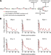
Strategy used to adapt SHIV-KNH1144 in rhesus macaques (RMs). (A) Serial passage of SHIV-KNH1144 in RMs. SHIV-KNH1144 was first passaged rapidly through a series of 6 naive RMs (RRp-9 through RHf-12). SHIV-KNH1144p1 was isolated from RHf-12 at week 2 postinoculation by cocultivation of RM PBMC. SHIV-KNH1144p1 was passaged further through RTp-9 (boxed), which was immunodepleted of CD8+ and CD20+ cells. Virus concentrated from plasma from RTp-9 at week 4 was designated SHIV-KNH1144p2 and used for next-generation sequencing. Afterwards, 2 naive RMs (RZy-11 and RZs-7) were inoculated via transfer of blood from RTp-9 at week 8 postinoculation, the time of necropsy. The biological isolate SHIV-KNH1144p4 was isolated from RZy-11 at week 36 postinoculation by RM PBMC coculture. w, week postinoculation. (B) Viral RNA (vRNA) loads of the first series of RMs in the rapid passage.
FIG 3
FIG 3
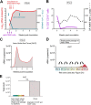
Virus passage in the immune-ablated RM RTp-9 depleted of CD8+ and B cells with cytotoxic MAbs. (A) RTp-9 was given doses of MAbs against CD8 (blue arrows) and CD20 (purple arrows) to destroy CD8+ T cells and NK cells as well as B cells, respectively. SHIV-KNH1144p1 was inoculated i.v. repeatedly (red arrows). vRNA loads and CD8+ T-cell counts are shown for RTp-9. The area under the curve (AUC) for vRNA loads is shown in gray. (B) Percentage of CD3CD19+ B cells (purple line) and CD4+ T-cell counts (dashed black line) in RTp-9. (C) AUC for vRNA load levels of RTp-9 over 8 weeks. (D) AUC for vRNA loads of each of the six RMs within the 2-week intervals between virus inoculation and transfer of infected blood to the next RM. (E) Sum of the AUC values for the initial 6 RMs (indicated by colors ranging from blue to red; panel D was compared to the AUC of the immunodepleted RM RTp-9 [gray]). 13×, 13-fold increase.
FIG 4
FIG 4
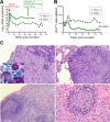
Virus passage in immunocompetent animals RZs-7 and RZy-11. (A) vRNA loads in RZs-7 and RZy-11. RZs-7 and RZy-11 were inoculated i.v. with 10 ml of blood from RTp-9 collected at necropsy. SHIV-KNH1144p4 was isolated 36 weeks after infection. (B) Peripheral CD4+ T-cell counts of RZs-7 (green line) and RZy-11 (dashed line). The dotted line (200 CD4+ T cells/μl) indicates severe depletion compatible with AIDS. (C) Histopathology of colon, lung, mesenteric lymph node, and liver in RM RZy-11 by H&E staining. Top left, the colonic lamina propria is markedly expanded by sheets of epithelioid macrophages, small numbers of lymphocytes, and plasma cells with loss of crypts (scale bar = 50 μm). Inset, acid-fast stain showing numerous intracytoplasmic acid-fast bacilli in macrophages (opportunistic infection). Top right, pulmonary granuloma composed of numerous epithelioid macrophages interspersed with lymphocytes and a multinucleated giant cell (arrowhead) (scale bar = 50 μm). Bottom left, mesenteric lymph node is multifocally infiltrated by numerous epithelioid macrophages (scale bar = 100 μm). Bottom right, the hepatic parenchyma contains a discrete granuloma composed of centrally located numerous epithelioid macrophages rimmed by small numbers of lymphocytes (scale bar = 20 μm).
FIG 5
FIG 5
Replication of adapted SHIV-KNH1144p4 in naive RM PBMC and coreceptor usage. (A) Replication of parental SHIV-KNH1144 and SHIV-KNH1144p4 in PBMC from 10 randomly selected naive RMs. The relatively lower level of replication in PBMC from donors 31334 and 31335 is shown at an appropriately lower scale in ng/ml (inset). (B) SHIV-KNH1144p4 was tested for usage of various coreceptors (CCR2, CCR3, CXCR4, CCR5, GPR15/BOB, and CXCR6/BONZO/STRL33) expressed on engineered U87.CD4 or Ghost cells.
FIG 6
FIG 6
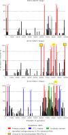
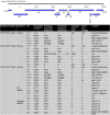
Frequency of mutations in the SHIV-KNH1144p1, SHIV-KNH1144p2, and SHIV-KNH1144p4 genomes. Sequence reads for each isolate were aligned to the parental SHIV-KNH1144. The height of each bar represents the percentage of sequence reads for a given mutation. Mutated reads at a prevalence of >5% are shown in the corresponding positions in the genome. Mutations identified in the SHIV-KNH1144p1 and SHIV-KNH1144p2 isolates emerged during adaptation in RMs without pressure from host adaptive immunity and are attributed to optimization of viral replication parameters (“fitness mutations”) in the new species; those observed at ≥80% prevalence are termed F1 to F9 and shown in red. Mutations observed in the biological isolate SHIV-KNH1144p4 occurred in the presence of host adaptive immunity, including CTL and Ab responses. Some fitness mutations had become fixed (F1 to F4 and F8) and are shown in red. Mutations appearing at ≥80% frequency are indicated in red (fitness mutations), blue (CTL-related mutations), or green (Ab-related mutations). Unique mutations appearing in the immunodepleted RM RTp-9 are highlighted yellow with a black box; F8 is highlighted yellow with no black box as it appears in the final isolate. Asterisks indicate that these mutations were also found in highly viremic RMs during the course of SIVmac239 infection (28).
FIG 7
FIG 7
Substitutions in SHIV-KNH1144p1, SHIV-KNH1144p2, and SHIV-KNH1144p4 compared with the parental SHIV-KNH1144. Footnote symbols: a, position in the parental SHIV-KNH1144 genome; b, convergent evolution resulting in optimized SIVmac239 sequences as described in reference 28; c, F4-1 and F4-2, the G→a mutation affects overlapping reading frames (underlined); an amino acid change occurred only in Rev. N/A, not applicable; C1, constant region 1; MSD, membrane-spanning domain; V2 or V3, variable loop 2 or 3; RT, reverse transcriptase; CTL, cytotoxic T lymphocyte; NHR, N-terminal heptad repeat. Mutated nucleotides are in lowercase letters.

References

    1. Hraber P, Korber BT, Lapedes AS, Bailer RT, Seaman MS, Gao H, Greene KM, McCutchan F, Williamson C, Kim JH, Tovanabutra S, Hahn BH, Swanstrom R, Thomson MM, Gao F, Harris L, Giorgi E, Hengartner N, Bhattacharya T, Mascola JR, Montefiori DC. 2014. Impact of clade, geography, and age of the epidemic on HIV-1 neutralization by antibodies. J Virol 88:12623–12643. doi:10.1128/JVI.01705-14. - DOI - PMC - PubMed
    1. Dowling WE, Kim B, Mason CJ, Wasunna KM, Alam U, Elson L, Birx DL, Robb ML, McCutchan FE, Carr JK. 2002. Forty-one near full-length HIV-1 sequences from Kenya reveal an epidemic of subtype A and A-containing recombinants. AIDS 16:1809–1820. doi:10.1097/00002030-200209060-00015. - DOI - PubMed
    1. Hemelaar J, Gouws E, Ghys PD, Osmanov S, WHO-UNAIDS Network for HIV Isolation and Characterisation . 2011. Global trends in molecular epidemiology of HIV-1 during 2000-2007. AIDS 25:679–689. doi:10.1097/QAD.0b013e328342ff93. - DOI - PMC - PubMed
    1. Rainwater S, DeVange S, Sagar M, Ndinya-Achola J, Mandaliya K, Kreiss JK, Overbaugh J. 2005. No evidence for rapid subtype C spread within an epidemic in which multiple subtypes and intersubtype recombinants circulate. AIDS Res Hum Retroviruses 21:1060–1065. doi:10.1089/aid.2005.21.1060. - DOI - PubMed
    1. Kiwanuka N, Laeyendecker O, Quinn TC, Wawer MJ, Shepherd J, Robb M, Kigozi G, Kagaayi J, Serwadda D, Makumbi FE, Reynolds SJ, Gray RH. 2009. HIV-1 subtypes and differences in heterosexual HIV transmission among HIV-discordant couples in Rakai, Uganda. AIDS 23:2479–2484. doi:10.1097/QAD.0b013e328330cc08. - DOI - PMC - PubMed

Publication types

MeSH terms

Substances