. 2019 Mar 5;116(10):4250-4255.
doi: 10.1073/pnas.1816556116. Epub 2019 Feb 13.

Dimers of mitochondrial ATP synthase induce membrane curvature and self-assemble into rows

Affiliations

Dimers of mitochondrial ATP synthase induce membrane curvature and self-assemble into rows

Thorsten B Blum et al. Proc Natl Acad Sci U S A. .

Abstract

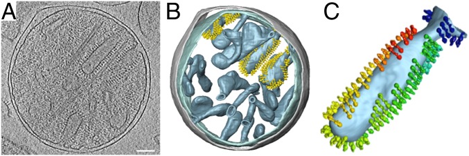
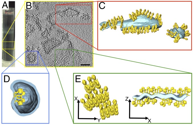
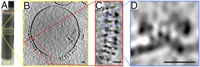
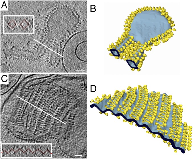
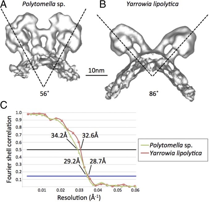
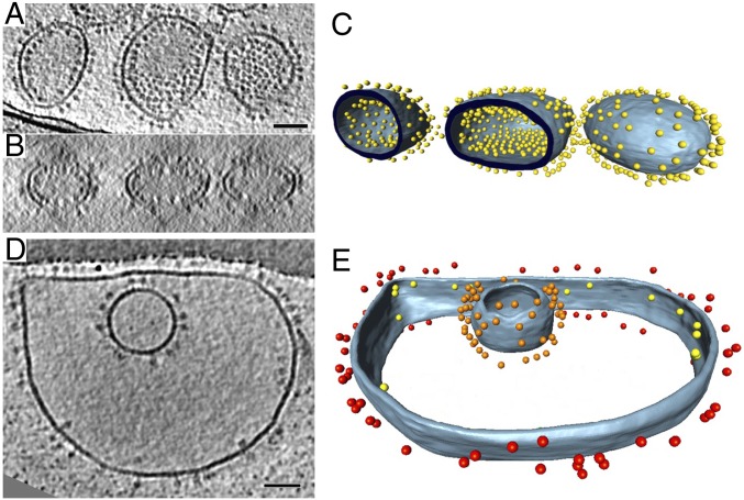
Mitochondrial ATP synthases form dimers, which assemble into long ribbons at the rims of the inner membrane cristae. We reconstituted detergent-purified mitochondrial ATP synthase dimers from the green algae Polytomella sp. and the yeast Yarrowia lipolytica into liposomes and examined them by electron cryotomography. Tomographic volumes revealed that ATP synthase dimers from both species self-assemble into rows and bend the lipid bilayer locally. The dimer rows and the induced degree of membrane curvature closely resemble those in the inner membrane cristae. Monomers of mitochondrial ATP synthase reconstituted into liposomes do not bend membrane visibly and do not form rows. No specific lipids or proteins other than ATP synthase dimers are required for row formation and membrane remodelling. Long rows of ATP synthase dimers are a conserved feature of mitochondrial inner membranes. They are required for cristae formation and a main factor in mitochondrial morphogenesis.

Keywords: ATP synthase; electron cryotomography; membrane curvature; mitochondria; subtomogram averaging.


Conflict of interest statement

The authors declare no conflict of interest.

Figures

Fig. 1.
Fig. 1.
Cryo-ET of isolated Polytomella sp. mitochondria. (A) Tomographic volume showing inner membrane cristae studded with ATP synthase dimers surrounded by the smooth, featureless outer membrane. (B) Segmented volumes show ATP synthase dimers arranged in rows. All cristae vesicles are similarly populated, but for clarity, ATP synthase dimers are shown in only three of them. Inner membrane, light blue; outer membrane, gray; ATP synthase dimers, yellow. (C) Individual club-shaped crista. Dimers in the membrane are replaced by subtomogram average volumes (rainbow colors). Dimer rows wrap around the tightly curved cristae edges. (Scale bar, 100 nm.)
Fig. 2.
Fig. 2.
Polytomella sp. ATP synthase dimers reconstituted at low lipid/protein ratio. (A) The lower gradient band (dashed yellow box) contains proteoliposomes with high protein density. (B) Cryo-ET indicates irregular vesicle shapes and membrane fragments densely packed with ATP synthase dimers. (Scale bar, 100 nm.) (CE) Segmented tomographic volumes. (C) Reconstituted ATP synthase dimers (yellow) form rows in the membrane (light blue). (D) Occasionally dimers insert with their F1 heads pointing into the vesicle, indicating that individual dimers impose local membrane curvature. (E) Membrane region with parallel ATP synthase dimer rows. The cross-section (Right) reveals that rows on opposite sides of the membrane distort the lipid bilayer into a corrugated sheet. C, D, and E are not on the same scale.
Fig. 3.
Fig. 3.
Polytomella sp. ATP synthase dimers reconstituted at high lipid/protein ratio. (A) The upper gradient band (yellow box) contains proteoliposomes with low protein density. (B) Cryo-ET of low-density vesicles indicates that the membrane is mostly empty and reconstituted dimers assemble into long rows (red box). (C) Consecutive dimers are laterally offset as in Polytomella cristae (Fig. 1C). (D) Cross-sections (blue box) reveal that rows bend the membrane locally. A subtomogram average of reconstituted dimer rows is shown in SI Appendix, Fig. S1. (Scale bars, 20 nm.)
Fig. 4.
Fig. 4.
Proteoliposomes of reconstituted Y. lipolytica ATP synthase dimers. (A and C) ATP synthase dimers (yellow) form rows that bend the lipid bilayer (light blue). Insets shows cross-sections (white lines), indicating that dimer rows bend the lipid bilayer by ∼90° (dashed red lines). (B) Flat membrane regions are devoid of ATP synthase. (D) Parallel rows of bidirectionally inserted dimers bend the bilayer into a corrugated sheet. (Scale bars, 100 nm.)
Fig. 5.
Fig. 5.
Subtomogram average volumes of ATP synthase dimers. (A) Polytomella (average of 103 dimer volumes). (B) Y. lipolytica (158 dimer volumes). Dimer angles between central stalks are indicated. (C) Gold-standard Fourier shell correlation (FSC) curves for the 3D reconstructions indicate a resolution of ∼29 Å at FSC = 0.143.
Fig. 6.
Fig. 6.
Liposomes reconstituted with Y. lipolytica ATP synthase monomers. (A and B) Tomographic slices of liposomes reconstituted with different concentrations of monomeric ATP synthases from Y. lipolytica. (C and E) Segmented volumes of A and D. Blue-gray, membrane; colored spheres, ATP synthases. (B) Colors represent ATP synthase F1 heads on different membrane surfaces. Red, outside of large outer vesicle; yellow, inside of large outer vesicles; orange, outside of small inner vesicle. Lipids are E. coli phosphatidyl ethanolamine (PE) 67%, phosphatidyl glycerol (PG) 23.2%, cardiolipin 9.8%. Percentages are in weight per volume. (Scale bar, 50 nm.)

References

    1. Kühlbrandt W. Structure and mechanisms of F-type ATP synthases. Annu Rev Biochem. 2019;88 in press. - PubMed
    1. Arnold I, Pfeiffer K, Neupert W, Stuart RA, Schägger H. Yeast mitochondrial F1F0-ATP synthase exists as a dimer: Identification of three dimer-specific subunits. EMBO J. 1998;17:7170–7178. - PMC - PubMed
    1. Davies KM, Anselmi C, Wittig I, Faraldo-Gómez JD, Kühlbrandt W. Structure of the yeast F1Fo-ATP synthase dimer and its role in shaping the mitochondrial cristae. Proc Natl Acad Sci USA. 2012;109:13602–13607. - PMC - PubMed
    1. Guo H, Bueler SA, Rubinstein JL. Atomic model for the dimeric FO region of mitochondrial ATP synthase. Science. 2017;358:936–940. - PMC - PubMed
    1. Lapaille M, et al. Atypical subunit composition of the chlorophycean mitochondrial F1FO-ATP synthase and role of Asa7 protein in stability and oligomycin resistance of the enzyme. Mol Biol Evol. 2010;27:1630–1644. - PubMed

Publication types

MeSH terms