. 2019 Feb 13;9(1):1971.
doi: 10.1038/s41598-018-37192-4.

Highly Efficient Transgenesis in Ferrets Using CRISPR/Cas9-Mediated Homology-Independent Insertion at the ROSA26 Locus

Affiliations

Highly Efficient Transgenesis in Ferrets Using CRISPR/Cas9-Mediated Homology-Independent Insertion at the ROSA26 Locus

Miao Yu et al. Sci Rep. .

Abstract

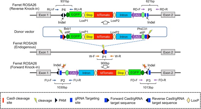
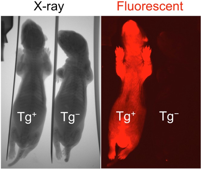
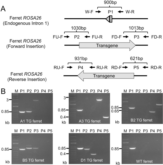
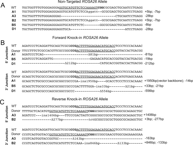
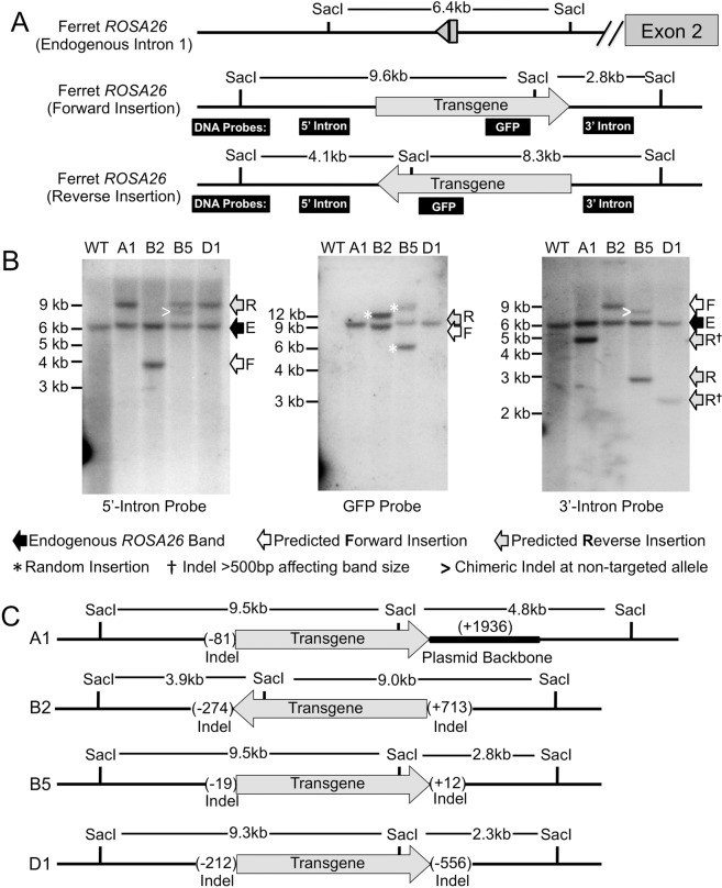
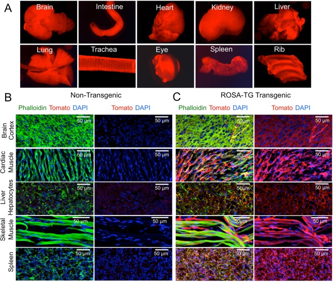
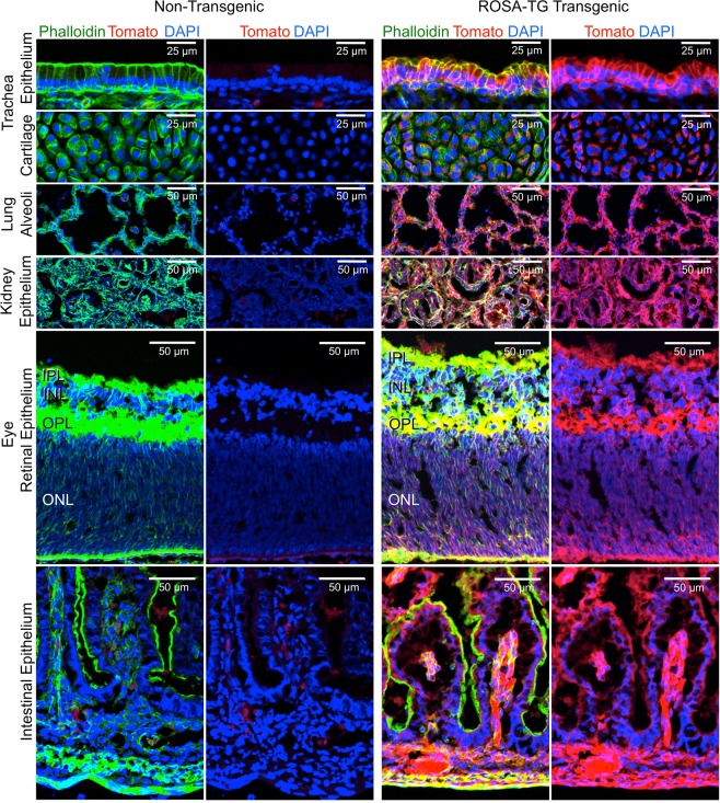
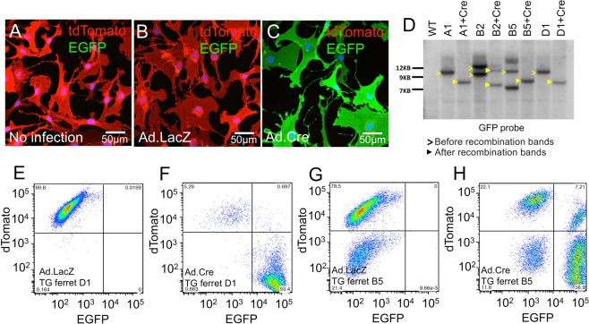
The domestic ferret (Mustela putorius furo) has proven to be a useful species for modeling human genetic and infectious diseases of the lung and brain. However, biomedical research in ferrets has been hindered by the lack of rapid and cost-effective methods for genome engineering. Here, we utilized CRISPR/Cas9-mediated, homology-independent insertion at the ROSA26 "safe harbor" locus in ferret zygotes and created transgenic animals expressing a dual-fluorescent Cre-reporter system flanked by PhiC31 and Bxb1 integrase attP sites. Out of 151 zygotes injected with circular transgene-containing plasmid and Cas9 protein loaded with the ROSA26 intron-1 sgRNA, there were 23 births of which 5 had targeted integration events (22% efficiency). The encoded tdTomato transgene was highly expressed in all tissues evaluated. Targeted integration was verified by PCR analyses, Southern blot, and germ-line transmission. Function of the ROSA26-CAG-LoxPtdTomatoStopLoxPEGFP (ROSA-TG) Cre-reporter was confirmed in primary cells following Cre expression. The Phi31 and Bxb1 integrase attP sites flanking the transgene will also enable rapid directional insertion of any transgene without a size limitation at the ROSA26 locus. These methods and the model generated will greatly enhance biomedical research involving lineage tracing, the evaluation of stem cell therapy, and transgenesis in ferret models of human disease.


Conflict of interest statement

The authors declare no competing interests.

Figures

Figure 1
Figure 1
Schematic strategy for targeted integration at the ferret ROSA26 locus and potential outcomes. Schematic shows the strategy of Cas9-mediated gene targeting and the donor vector. The transgene cassette of the donor plasmid contains a CAG promoter with intron driving expression of the LoxPtdTomatoStopLoxPEGFP-PolyA transgene with flanking Bxb1/PhiC31 attP integrase sites. The strategy incorporates the Cas9 gRNA sequences targeting the ROSA26 intron 1 into the flanking sequence of the donor plasmid. After injection of the Cas9 RNP complex into zygote pronuclei, the donor plasmid and chromosome are synchronously cleaved, allowing for efficient integration of the donor DNA. Integration is expected to occur more frequently in the reverse orientation since the gRNA targeting sequence remains intact in the forward insertion if indels are not generated. P1–5 indicate the location of primer pairs for identification the integration junctions and orientations. Unlabeled annotations used in this figure are denoted in the box.
Figure 2
Figure 2
Whole body tdTomato expression in a transgenic ferret. tdTomato transgene expression is shown for a juvenile F1 transgene positive ferret founder A1 (Tg+) and transgene negative littermate (Tg). X-ray images are shown in the left panel and fluorescent images are shown in the right panel.
Figure 3
Figure 3
Genotyping of transgene integration and orientation by PCR analysis. To map the orientation of the transgene integration events at the ROSA26 locus and to characterize indels at the 5′ and 3′ junctions of the insertion site, genomic DNA from F0 founders with observed en face tdTomato expression and non-transgenic wild-type ferrets were analyzed for transgene integration using a PCR assay. (A) Schematic of the PCR strategy and primers used for mapping the direction of integration events. (B) Representative gel images of PCR products (P1-P5) for the indicated transgenic (TG) founder ferrets and a non-transgenic wild-type (WT) animal (see Table 2 for primer descriptions). The predicted sizes of PCR products varied among distinct TG ferret founders due to different sized indels generated from error-prone NHEJ-mediated integration. The sizes of these indels were confirmed by Sanger sequencing of the PCR products (Fig. 4). M, lane denotes 1 kb plus marker.
Figure 4
Figure 4
Characterization of indels at the transgene integration sites. The PCR-specific amplicons as shown in Fig. 3 were gel extracted and analyzed by Sanger sequencing. (A) Indels created at the non-targeted ROSA26 locus for the indicated founder animals. (B,C) Indels created at the targeted ROSA26 locus for (B) forward and (C) reverse integration events. Wild-type and donor sequences are shown at the top with the targeting gRNA site underlined and protospacer adjacent motif (PAM) sequences bolded. The dash lines denote deletions (Δ) and insertions (ins) with the number of nucleotides indicated. Italics font denotes mutations at the indel site.
Figure 5
Figure 5
Southern blot analysis of transgenic founder genomic DNA. (A) Schematic drawing of hybridization probes for the external arms of 3′-intron and 5′-intron and the EGFP transgene, with predicted sizes of Sac1 digested bands for the non-targeted and forward- and reverse-integrated transgenes, respectively. (B) Representative Sac1-restricted Southern blots following hybridization with the indicated probes for the four surviving transgenic ferrets. Arrows to the left of each blot denote bands for the endogenous intact locus (E), forward (F)- and reverse (R)-orientated insertion of the transgene cassette. The extra bands seen in B2 and B5 transgenic ferrets with the EGFP probe (white asterisks) are suggestive of a random integration event in these animals. Other annotations on the blots are marked in the legend at the bottom. (C) Schematic showing the predicted transgene orientation and size of indels at both the 5′ and 3′ junctions of the transgene insertion site for the indicated transgenic animals as determined by results of sequencing and Southern blotting.
Figure 6
Figure 6
Global expression of the encoded tdTomato transgene in ROSA26-targeted (ROSA-TG) ferrets. (A) Representative en face fluorescent images of the indicated whole mount organs in the transgenic F1 newborn ferret (A1) demonstrate ubiquitous expression of the tdTomato transgene. (B,C) Fluorescent photomicrographs of phalloidin-stained (green) cryosections for brain cortex, cardiac muscle, liver hepatocytes, skeletal muscle and spleen from a newborn (B) non-transgenic and (C) transgenic ferret (A1). Phalloidin was used for staining filamentous actin (F-actin) to allow for better visualization of tissue structure. DAPI was used to stain nuclei.
Figure 7
Figure 7
Global expression of the encoded tdTomato transgene in epithelial tissues of ROSA26-targeted (ROSA-TG) ferrets. Fluorescent photomicrographs of phalloidin-stained (green) cryosections of the indicated tissues including tracheal epithelium, tracheal cartilage, distal lung alveoli, kidney epithelium, retinal epithelium, and intestinal epithelium in a non-transgenic (left panels) and a transgenic F1 (A1; right panels) newborn ferret. DAPI was used to stain nuclei. INL, inner nuclear layer; IPL, inner plexiform layer; ONL, outer nuclear layer; OPL, outer plexiform layer. Phalloidin was used for staining filamentous actin (F-actin) to allow for better visualization of tissue structure. DAPI was used to stain nuclei.
Figure 8
Figure 8
Functional analysis of Cre/LoxP-mediated recombination in fibroblast from ROSA26-targeted transgenic ferrets. Passage 4 (P4) primary fibroblasts derived from all surviving transgenic ferret founders were used for functional analysis of Cre-mediated recombination in vitro. (AC) In vitro functional validation of Cre-mediated recombination in transgenic ferret fibroblasts (A1). Representative confocal fluorescent images for tdTomato and EGFP of the (A) mock control non-infected fibroblasts, (B) negative control Ad.LacZ-infected fibroblasts, and (C) experimental Ad.Cre-infected fibroblasts at 14 days post-infection. (D) Southern blotting analysis of Ad.Cre-infected transgenic ferret fibroblasts. Fibroblasts from the surviving A1, B2, B5 and D1 transgenic ferrets were infected with Ad.Cre and genomic DNA was extracted for Sac1-restricted Southern blotting against an EGFP probe at 14 days post-infection. Wild-type non-transgenic ferret DNA and non-infected fibroblasts of each transgenic animal served as negative controls for Cre-mediated excision of tdTomato. Open and closed arrowheads on the blot indicate the intact and tdTomato-deleted transgene fragment, respectively. (EH) Representative FACS plots of tdTomato and EGFP fluorescence following Cre-mediated recombination in fibroblast cells from transgenic ferrets. Fibroblasts derived from transgenic ferret founders D1 (E,F) and B5 (G,H) were analyzed at 14 days after an infection of Ad.LacZ (E,G) and Ad.Cre (FH). No EGFP conversion was detected by FACS in Ad.LacZ-infected fibroblasts derived from D1 (E) and B5 (G) animals, but striking conversions of tdTomato to EGFP were observed in cells infected with Ad.Cre from both transgenic founders D1 (F) and B5 (H). The two other transgenic founder lines (A1 and B2) demonstrated a FACS pattern following Ad.Cre infection similar to D1. Note that B5 fibroblasts has a subset of tdTomato negative cells in both Ad.Cre and Ad.LacZ infected cultures, suggesting this founder is a chimera with integration at the ROSA26 locus occurring after division of the zygote. Values in the corners of the FACS plots indicate the percentage of cells in each quadrant.

References

    1. Sun X, et al. Disease phenotype of a ferret CFTR-knockout model of cystic fibrosis. J Clin Invest. 2010;120:3149–3160. doi: 10.1172/JCI43052. - DOI - PMC - PubMed
    1. Sun X, et al. Adeno-associated virus-targeted disruption of the CFTR gene in cloned ferrets. J Clin Invest. 2008;118:1578–1583. doi: 10.1172/JCI34599. - DOI - PMC - PubMed
    1. Keiser NW, et al. Defective innate immunity and hyperinflammation in newborn cystic fibrosis transmembrane conductance regulator-knockout ferret lungs. Am J Respir Cell Mol Biol. 2015;52:683–694. doi: 10.1165/rcmb.2014-0250OC. - DOI - PMC - PubMed
    1. Johnson MB, et al. Aspm Knockout Ferret Reveals an Evolutionary Mechanism Governing Cerebral Cortical Size. Nature. 2018;556:370–375. doi: 10.1038/s41586-018-0035-0. - DOI - PMC - PubMed
    1. Enkirch T, von Messling V. Ferret models of viral pathogenesis. Virology. 2015;479–480:259–270. doi: 10.1016/j.virol.2015.03.017. - DOI - PMC - PubMed

Publication types

MeSH terms

Substances