. 2019 Feb 13;9(1):1881.
doi: 10.1038/s41598-018-38325-5.

Tumorigenicity assay essential for facilitating safety studies of hiPSC-derived cardiomyocytes for clinical application

Affiliations

Tumorigenicity assay essential for facilitating safety studies of hiPSC-derived cardiomyocytes for clinical application

Emiko Ito et al. Sci Rep. .

Abstract

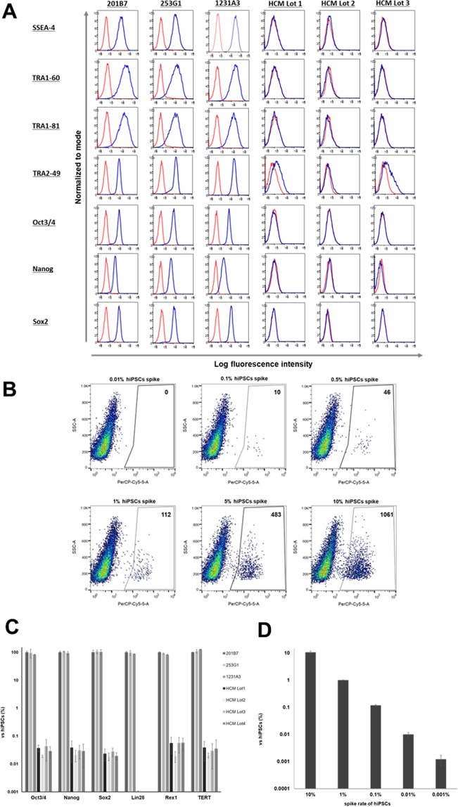
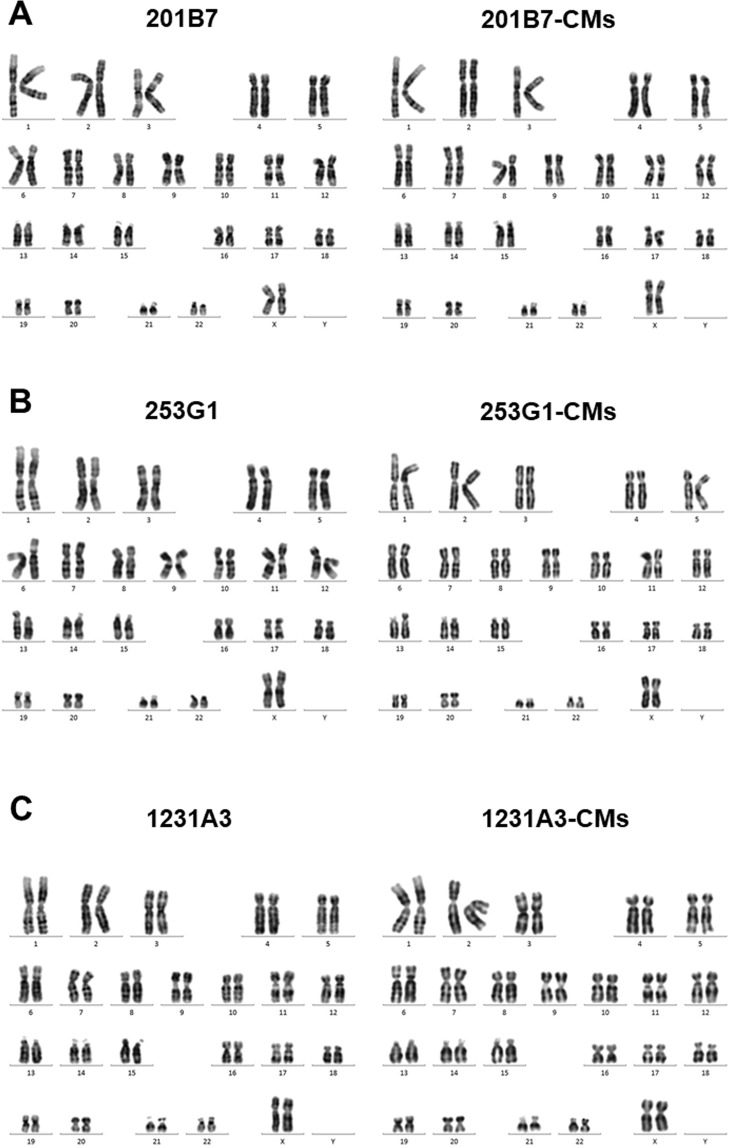
Transplantation of cardiomyocytes (CMs) derived from human induced pluripotent stem cells (hiPSC-CMs) is a promising treatment for heart failure, but residual undifferentiated hiPSCs and malignant transformed cells may lead to tumor formation. Here we describe a highly sensitive tumorigenicity assay for the detection of these cells in hiPSC-CMs. The soft agar colony formation assay and cell growth analysis were unable to detect malignantly transformed cells in hiPSC-CMs. There were no karyotypic abnormalities during hiPSCs subculture and differentiation. The hiPSC markers TRA1-60 and LIN28 showed the highest sensitivity for detecting undifferentiated hiPSCs among primary cardiomyocytes. Transplantation of hiPSC-CMs with a LIN28-positive fraction > 0.33% resulted in tumor formation in nude rats, whereas no tumors were formed when the fraction was < 0.1%. These findings suggested that combination of these in vitro and in vivo tumorigenecity assays can verify the safety of hiPSC-CMs for cell transplantation therapy.


Conflict of interest statement

The authors declare no competing interests.

Figures

Figure 1
Figure 1
Differentiation of human iPSCs into hiPSCs-CMs in vitro. (A) Representative flow cytometry data for hiPSCs-CMs labeled with anti-cTnT antibody. (B) mRNA expression of aMHC and cTNT in hiPSC-CMs as compared to hiPSCs as determined by qRT-PCR. **P < 0.01. (C) Immunolabeling of hiPSC-CMs with anti-cTNT (green) and anti-sarcomeric α-actinin (red) antibodies with Hoechst 33342 staining. Scale bar, 50 μm.
Figure 2
Figure 2
Detection of malignantly transformed cells in vitro. (AC) Soft agar colony formation assay. Phase contrast micrographs of 201B7 cells, 201B7-CMs, human primary cardiomyocytes (HCM), and HeLa cells spiked into HCM (1%) cultured in soft agar medium for (A) 10 days and (B) 20 days. Arrows indicate colonies. (C) Quantitative analysis of fluorescence reflecting colony formation in hiPSCs, hiPSC-CMs, HCM Lot2~4, and HeLa cells spiked into HCM Lot1. Experiments were performed in triplicate. Each bar represents mean ± SD. **P < 0.01 vs. the 0% control. (D,E) Analysis of hiPSCs, hiPSCs-CMs, and HeLa cell growth and growth curves of hMSC contaminated with HeLa cells (D) and hiPSC-CMs (E).
Figure 3
Figure 3
Detection of undifferentiated hiPSCs in vitro. (A,B) Flow cytometry analysis of hiPSCs and primary cardiomyocytes in vitro. (A) Flow cytometry data of hiPSCs and primary cardiomyocytes labeled with antibodies against a variety of stem cell markers including Oct3/4, Nanog, Sox2, SSEA-4, TRA1-60, TRA1-81, and TRA2-49. (B) 201B7 cells were spiked into primary cardiomyocytes at concentrations of 10%, 5%, 1%, 0.5%, 0.1%, and 0.01% and analyzed by flow cytometry with anti-TRA 1-60 antibody. (C) mRNA expression levels of Oct3/4, Nanog, Sox2, Rex1, and telomerase reverse transcriptase (TERT) in hiPSCs and primary cardiomyocytes, as detected by qRT-PCR. (D) 201B7 cells were spiked into primary cardiomyocytes at concentrations of 10%, 1%, 0.1%, 0.01%, and 0.001% and LIN28 mRNA level was evaluated by qRT-PCR.
Figure 4
Figure 4
Karyotype analysis. Representative karyograms of (A) 201B7 cells and 201B7-CMs, (B) 253G1 cells and 201B7-CMs, (C) 1231A3 cells and 1231A3-CMs.
Figure 5
Figure 5
Detection of undifferentiated hiPSCs in vivo. Transplantation of hiPSC-CM sheets into the left ventricular surface of immunodeficient rats. (A) Representative H&E staining of teratoma. (B) Relationship between LIN28 mRNA expression in hiPSC-CMs by cell line and tumor formation. (C) Relationship between LIN28 mRNA expression in hiPSC-CMs and tumor formation. (D) ROC curves for LIN28 mRNA expression in all hiPSC-CMs and tumor formation.

References

    1. Yoshida Y, Yamanaka S. iPS cells: a source of cardiac regeneration. J. Mol. Cell. Cardiol. 2011;50:327–332. doi: 10.1016/j.yjmcc.2010.10.026. - DOI - PubMed
    1. Yoshida Y, Yamanaka S. Induced pluripotent stem cells 10 years later: for cardiac applications. Circ. Res. 2017;120:1958–1968. doi: 10.1161/CIRCRESAHA.117.311080. - DOI - PubMed
    1. Miyagawa S. Translational research in iPS cell derived cardiomyocyte sheet for the treatment of heart failure. Nihon Rinsho. 2016;74:667–70. - PubMed
    1. Takahashi K, et al. Induction of pluripotent stem cells from adult human fibroblasts by defined factors. Cell. 2007;131:861–72. doi: 10.1016/j.cell.2007.11.019. - DOI - PubMed
    1. Sougawa N. et al. Immunologic targeting of CD30 eliminates tumourigenic human pluripotent stem cells, allowing safer clinical application of hiPSC-based cell therapy. Sci Rep. Published online February 27, 2018, 10.1038/s41598-018-21923-8 (2018). - PMC - PubMed

Publication types