Review
. 2019 Jan 25:13:1.
doi: 10.3389/fnana.2019.00001. eCollection 2019.

Applications of Light-Sheet Microscopy in Microdevices

Affiliations
Review

Applications of Light-Sheet Microscopy in Microdevices

Ignacio Albert-Smet et al. Front Neuroanat. .

Abstract

Light-sheet fluorescence microscopy (LSFM) has been present in cell biology laboratories for quite some time, mainly as custom-made systems, with imaging applications ranging from single cells (in the micrometer scale) to small organisms (in the millimeter scale). Such microscopes distinguish themselves for having very low phototoxicity levels and high spatial and temporal resolution, properties that make them ideal for a large range of applications. These include the study of cellular dynamics, in particular cellular motion which is essential to processes such as tumor metastasis and tissue development. Experimental setups make extensive use of microdevices (bioMEMS) that provide better control over the substrate environment than traditional cell culture experiments. For example, to mimic in vivo conditions, experiment biochemical dynamics, and trap, move or count cells. Microdevices provide a higher degree of empirical complexity but, so far, most have been designed to be imaged through wide-field or confocal microscopes. Nonetheless, the properties of LSFM render it ideal for 3D characterization of active cells. When working with microdevices, confocal microscopy is more widespread than LSFM even though it suffers from higher phototoxicity and slower acquisition speeds. It is sometimes possible to illuminate with a light-sheet microdevices designed for confocal microscopes. However, these bioMEMS must be redesigned to exploit the full potential of LSFM and image more frequently on a wider scale phenomena such as motion, traction, differentiation, and diffusion of molecules. The use of microdevices for LSFM has extended beyond cell tracking studies into experiments regarding cytometry, spheroid cultures and lab-on-a-chip automation. Due to light-sheet microscopy being in its early stages, a setup of these characteristics demands some degree of optical expertise; and designing three-dimensional microdevices requires facilities, ingenuity, and experience in microfabrication. In this paper, we explore different approaches where light-sheet microscopy can achieve single-cell and subcellular resolution within microdevices, and provide a few pointers on how these experiments may be improved.

Keywords: SPIM; cellular imaging; light-sheet fluorescence microscopy; microdevices; microfluidics.


Figures

Figure 1
Figure 1
Schematic of Fluorescence Light-Sheet Microscopy techniques. The figure shows illustrations of the different LSFM techniques for imaging in microdevices. (A) Titled LSFM; (B) Oblique Plane Microscopy; (C) Inverted SPIM; (D) Dual-illumination iSPIM; (E) Open-top SPIM; (F) Diagonal SPIM; (G) Reflected SPIM; (H) Single-objective SPIM. In the diagrams, the blue arrows denote the direction of excitation light traveling the objective, while the green arrows show the path of emission light. The light-sheet created is shown in light blue, and focuses in the specimen (a cell) within a microfluidic device supported on a coverslip. In Oblique Plane Microscopy (B) the size of the objective is disproportionally large to represent that an objective of high numerical aperture is required.
Figure 2
Figure 2
Comparison between the inverted SPIM and open-top SPIM configurations. Illustrations of iSPIM and open-top SPIM are shown imaging a cell in a PDMS microfluidic device with some bubbles. The blue arrows denote excitation light, while the green arrows show the path of emission light. In iSPIM, lighter blue and lighter green arrows show the reflectance caused by bubbles, which is avoided in open-top SPIM by imaging through the refractive medium prism and the glass coverslip. The working distance (WD) of both setups is illustrated, exemplifying another of the advantages of open-top SPIM which allows objectives of shorted working distance which are generally those of higher numerical aperture and magnification.
Figure 3
Figure 3
Single-objective LSFM microfluidic chip layout [Figure 6 from Meddens et al. (2016)]. (A) Exploded diagram of the different PMMA layers packaging the microfluidic chip. (B) Top and side view images of the assembled chip. (C) Schematic of the microfluidic device showing channel connections. (D) Photograph of the top of the microdevice. (E) Photograph of the bottom of the microdevice sealed with a coverslip.
Figure 4
Figure 4
Mirror-embedded microfluidic chip for single-objective plane illumination [Figure 3 from Miura et al. (2018)]. (A) Schematic of the channels in the microfluidic device. (i) Hydrodynamic focusing region. (ii) Optical interrogation region. (B) Diagram summarizing the microfabrication process. (C) Photograph of the microfluidic chip. (D) Close-up photograph of the microchannel and aluminum-coated reflective glass.
Figure 5
Figure 5
Design of a microfluidic chip with integrated waveguide for on-chip light-sheet illumination [Figure 1 from Deschout et al. (2014)]. Laser light (green) from a fiber optic enters the planar waveguide and is confined in the vertical direction, but spreads horizontally forming a sheet of light that exits in the microchannel. The fluorescence light (red) is collected by an objective lens. The red dots show fluorophores that are excited by the light-sheet, while black dots are those that are not. Only the fluorophore molecules traversed by the light-sheet are excited, removing out of focus noise.
Figure 6
Figure 6
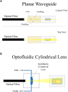
On-chip plane illumination methods based on optical fiber delivery: (A) A microfabricated planar waveguide allows the light from the optical fiber to expand laterally but not vertical. Confining the light in such a way creates an illumination plane which emerges at the microchannel where the sample is placed. (B) In this case, an optofluidic cylindrical lens creates a plane of light from an incident beam. The focus of such cylindrical lens can be changed if liquids of different refractive indices are employed. Two microchannels connect to the cylindrical microfabricated gap to change the fluid.
Figure 7
Figure 7
Compact, rapid, and cost-effective optofluidic device capable of preparing customized sample droplets [Figure 1 from Jiang et al. (2017)]. (A) Diagram of the device structure that includes the illumination optics, a microfluidic chip and a 3D-printed base. (B) The working scheme of the flow-based light-sheet scanning of droplets. The functions of droplet formation, mixing, LSFM imaging and bypassing are sequentially integrated, from upstream to downstream. Imaging is performed by the microscope objective from the side facet of the microfluidic device. (C) The device can be mounted on an inverted microscope for sustained image acquisition. Flow-based scanning eliminates the need of motorized stage and increases throughput. (D) The pictures of the actual droplets traveling through the microchannels.

References

    1. Ancora D., Di Battista D., Giasafaki G., Psycharakis S. E., Liapis E., Ripoll J., et al. (2017). Phase-retrieved tomography enables mesoscopic imaging of opaque tumor spheroids. Sci. Rep. 7:11854. 10.1038/s41598-017-12193-x - DOI - PMC - PubMed
    1. Arranz A., Dong D., Zhu S., Rudin M., Tsatsanis C., Tian J., et al. (2013). Helical optical projection tomography. Opt. Express 21 25912–25925. 10.1364/OE.21.025912 - DOI - PubMed
    1. Bouchard M. B., Voleti V., Mendes C. S., Lacefield C., Grueber W. B., Mann R. S., et al. (2015). Swept confocally-aligned planar excitation (SCAPE) microscopy for high-speed volumetric imaging of behaving organisms. Nat. Photonics 9 113–119. 10.1038/nphoton.2014.323 - DOI - PMC - PubMed
    1. Bruns T., Schickinger S., Wittig R., Schneckenburger H. (2012). Preparation strategy and illumination of three-dimensional cell cultures in light sheet–based fluorescence microscopy. J. Biomed. Opt. 17:1015181. 10.1117/1.JBO.17.10.101518 - DOI - PubMed
    1. Chen B. C., Legant W. R., Wang K., Shao L., Milkie D. E., Davidson M. W., et al. (2014). Lattice light-sheet microscopy: imaging molecules to embryos at high spatiotemporal resolution. Science 346:1257998. 10.1126/science.1257998 - DOI - PMC - PubMed