

Schisandrin A Inhibits the IL-1β-Induced Inflammation and Cartilage Degradation via Suppression of MAPK and NF-κB Signal Pathways in Rat Chondrocytes
- PMID: 30761007
- PMCID: PMC6361757
- DOI: 10.3389/fphar.2019.00041
Schisandrin A Inhibits the IL-1β-Induced Inflammation and Cartilage Degradation via Suppression of MAPK and NF-κB Signal Pathways in Rat Chondrocytes
Abstract
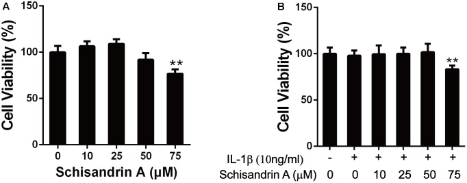
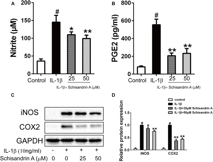
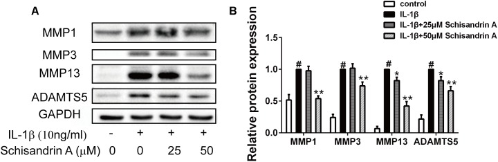
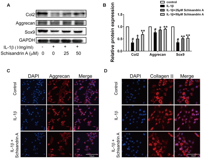
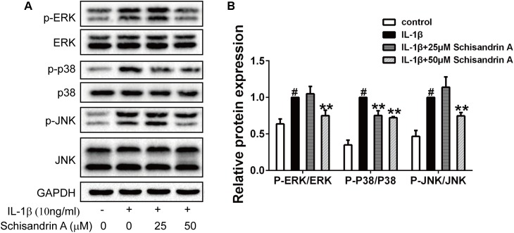
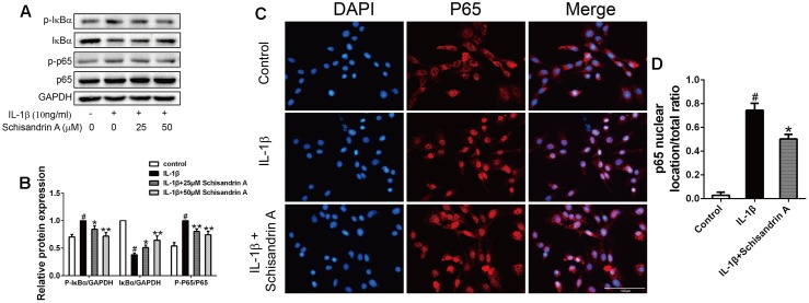
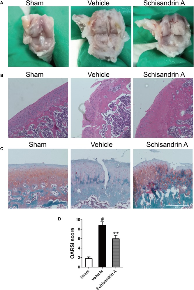
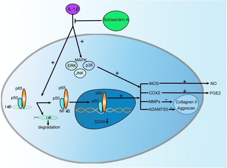
Osteoarthritis (OA) is a common joint disease in the elderly population. Its development has been reported to be associated with cartilage degradation and inflammatory responses. Schisandrin A, a bioactive lignin in Schisandra sphenanthera, has shown its anti-inflammatory potential in various inflammation diseases. However, the effects of Schisandrin A on OA remain to explore. In this study, rat chondrocytes were treated with IL-1β (10 ng/ml) with or without different concentrations of Schisandrin A for 24 h. Cell viability was evaluated by CCK-8 assay. Production of nitric oxide (NO) and prostaglandin E2 (PGE2) was measured by the Griess reaction and ELISA. The MAPK/NF-κB-related signaling molecules expression and the protein production of inducible nitric oxide synthase (iNOS), cyclooxygenase (Cox)-2, MMPs (MMP1, MMP3, MMP13), ADAMTS5, Collagen II, aggrecan, and Sox9 were detected by Western blot. Protein expression of Collagen II, aggrecan, and p65 nuclear translocation was evaluated by immunofluorescence. In vivo, intra-articular injection of 50 μM Schisandrin A or equal volume of vehicle was performed on rat OA models. Severity of cartilage damage was evaluated by HE and Safranin-O-Fast green staining. Our results revealed that Schisandrin A could suppress the IL-1β-induced production of NO and PGE2 in rat chondrocytes. Consistent with these findings, the upregulation of iNOS and Cox2 could also been decreased by Schisandrin A. Additionally, Schisandrin A could inhibit IL-1β-induced cartilage matrix catabolic enzymes including MMPs and ADAMTS5. Moreover, the IL-1β-induced downregulation of Collagen II, aggrecan, and Sox9 could be ameliorated by Schisandrin A. Mechanistically, Schisandrin A functioned by suppressing MAPK and NF-κB signal pathways. In vivo, Schisandrin A prevented cartilage damage in rat OA model. In conclusion, this study elucidates that Schisandrin A inhibits the IL-1β-induced inflammation and cartilage degradation via suppression of MAPK and NF-κB signal pathways, indicating its potential role in OA therapy.
Keywords: ADAMTS5; Cox-2; MAPK; MMPs; NF-κB; Schisandrin A; iNOS; osteoarthritis.
Figures
Similar articles
-
Schisandrin B ameliorated chondrocytes inflammation and osteoarthritis via suppression of NF-κB and MAPK signal pathways.Drug Des Devel Ther. 2018 May 9;12:1195-1204. doi: 10.2147/DDDT.S162014. eCollection 2018. Drug Des Devel Ther. 2018. PMID: 29785089 Free PMC article.
-
Neferine Inhibits Expression of Inflammatory Mediators and Matrix Degrading Enzymes in IL-1β-Treated Rat Chondrocytes via Suppressing MAPK and NF-κB Signaling Pathways.Inflammation. 2020 Aug;43(4):1209-1221. doi: 10.1007/s10753-019-01143-6. Inflammation. 2020. PMID: 32488683
-
Alpha-Mangostin protects rat articular chondrocytes against IL-1β-induced inflammation and slows the progression of osteoarthritis in a rat model.Int Immunopharmacol. 2017 Nov;52:34-43. doi: 10.1016/j.intimp.2017.08.010. Epub 2017 Aug 31. Int Immunopharmacol. 2017. PMID: 28858724
-
Efficacy of some Herbal Medicines in Osteoarthritis with a Focus on Topical Agents: A Systematic Review.Curr Pharm Des. 2020;26(22):2676-2681. doi: 10.2174/1381612826666200429013728. Curr Pharm Des. 2020. PMID: 32348208
-
ADAMTS5 in Osteoarthritis: Biological Functions, Regulatory Network, and Potential Targeting Therapies.Front Mol Biosci. 2021 Aug 9;8:703110. doi: 10.3389/fmolb.2021.703110. eCollection 2021. Front Mol Biosci. 2021. PMID: 34434966 Free PMC article. Review.
Cited by
-
Curculigoside inhibits osteoarthritis <em>via</em> the regulation of NLRP3 pathway.Eur J Histochem. 2023 Dec 13;67(4):3896. doi: 10.4081/ejh.2023.3896. Eur J Histochem. 2023. PMID: 38112591 Free PMC article.
-
Downregulation of microRNA-23b-3p alleviates IL-1β-induced injury in chondrogenic CHON-001 cells.Drug Des Devel Ther. 2019 Jul 23;13:2503-2512. doi: 10.2147/DDDT.S211051. eCollection 2019. Drug Des Devel Ther. 2019. PMID: 31440033 Free PMC article.
-
Cardamonin Inhibited IL-1β Induced Injury by Inhibition of NLRP3 Inflammasome via Activating Nrf2/NQO-1 Signaling Pathway in Chondrocyte.J Microbiol Biotechnol. 2021 Jun 28;31(6):794-802. doi: 10.4014/jmb.2103.03057. J Microbiol Biotechnol. 2021. PMID: 34024891 Free PMC article.
-
Tissue-specific transcriptome and metabolome analyses reveal candidate genes for lignan biosynthesis in the medicinal plant Schisandra sphenanthera.BMC Genomics. 2023 Oct 11;24(1):607. doi: 10.1186/s12864-023-09628-3. BMC Genomics. 2023. PMID: 37821824 Free PMC article.
-
Midazolam as a Probe for Heterotropic Drug-Drug Interactions Mediated by CYP3A4.Biomolecules. 2022 Jun 20;12(6):853. doi: 10.3390/biom12060853. Biomolecules. 2022. PMID: 35740978 Free PMC article.
References
LinkOut - more resources
Full Text Sources
Research Materials
Miscellaneous