

Ventilatory Limitation of Exercise in Pediatric Subjects Evaluated for Exertional Dyspnea
- PMID: 30761012
- PMCID: PMC6361738
- DOI: 10.3389/fphys.2019.00020
Ventilatory Limitation of Exercise in Pediatric Subjects Evaluated for Exertional Dyspnea
Abstract
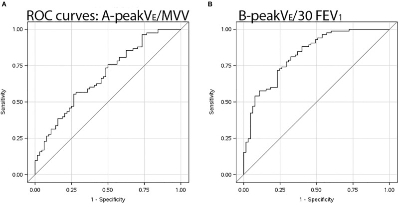
Purpose: Attribution of ventilatory limitation to exercise when the ratio of ventilation ( ) at peak work to maximum voluntary ventilation (MVV) exceeds 0.80 is problematic in pediatrics. Instead, expiratory flow limitation (EFL) measured by tidal flow-volume loop (FVL) analysis - the method of choice - was compared with directly measured MVV or proxies to determine ventilatory limitation. Methods: Subjects undergoing clinical evaluation for exertional dyspnea performed maximal exercise testing with measurement of tidal FVL. EFL was defined when exercise tidal FVL overlapped at least 5% of the maximal expiratory flow-volume envelope for > 5 breaths in any stage of exercise. We compared this method of ventilatory limitation to traditional methods based on MVV or multiples (30, 35, or 40) of FEV1. Receiver operating characteristic curves were constructed and area under curve (AUC) computed for peak /MVV and peak /x⋅FEV1. Results: Among 148 subjects aged 7-18 years (60% female), EFL was found in 87 (59%). Using EFL shown by FVL analysis as a true positive to determine ventilatory limitation, AUC for peak /30⋅FEV1 was 0.84 (95% CI 0.78-0.90), significantly better than AUC 0.70 (95% CI 0.61-0.79) when 12-s sprint MVV was used for peak /MVV. Sensitivity and specificity were 0.82 and 0.70 respectively when using a cutoff of 0.85 for peak /30⋅FEV1 to predict ventilatory limitation to exercise. Conclusion: Peak /30⋅FEV1 is superior to peak /MVV, as a means to identify potential ventilatory limitation in pediatric subjects when FVL analysis is not available.
Keywords: children; dyspnea; exercise; flow limitation; flow-volume curve; ventilation.
Figures
Similar articles
-
Calculated versus Measured MVV-Surrogate Marker of Ventilatory Capacity in Pediatric CPET.Med Sci Sports Exerc. 2017 Oct;49(10):1987-1992. doi: 10.1249/MSS.0000000000001318. Med Sci Sports Exerc. 2017. PMID: 28489684
-
Tidal Flow-Volume Loop Enveloping at Rest in Advanced COPD.Respir Care. 2019 Dec;64(12):1488-1499. doi: 10.4187/respcare.06787. Epub 2019 Aug 27. Respir Care. 2019. PMID: 31455685
-
Emerging concepts in the evaluation of ventilatory limitation during exercise: the exercise tidal flow-volume loop.Chest. 1999 Aug;116(2):488-503. doi: 10.1378/chest.116.2.488. Chest. 1999. PMID: 10453881 Review.
-
Ventilatory capacity and its utilisation during exercise.Lung. 2008 Sep-Oct;186(5):345-50. doi: 10.1007/s00408-008-9101-y. Epub 2008 Jul 3. Lung. 2008. PMID: 18597141
-
Flow limitation: an overview.Monaldi Arch Chest Dis. 1999 Aug;54(4):353-7. Monaldi Arch Chest Dis. 1999. PMID: 10546481 Review.
Cited by
-
Cardiopulmonary Exercise Performance of Children Born Non-Extremely Preterm.Children (Basel). 2024 Feb 4;11(2):198. doi: 10.3390/children11020198. Children (Basel). 2024. PMID: 38397309 Free PMC article.
-
Pitfalls in Expiratory Flow Limitation Assessment at Peak Exercise in Children: Role of Thoracic Gas Compression.Med Sci Sports Exerc. 2020 Nov;52(11):2310-2319. doi: 10.1249/MSS.0000000000002378. Med Sci Sports Exerc. 2020. PMID: 33064406 Free PMC article.
-
Exercise Limitation in Children and Adolescents with Mild-to-Moderate Asthma.J Asthma Allergy. 2022 Jan 18;15:89-98. doi: 10.2147/JAA.S335357. eCollection 2022. J Asthma Allergy. 2022. PMID: 35082501 Free PMC article.
References
LinkOut - more resources
Full Text Sources