

Intestinal Microbiota as a Host Defense Mechanism to Infectious Threats
- PMID: 30761120
- PMCID: PMC6362409
- DOI: 10.3389/fmicb.2018.03328
Intestinal Microbiota as a Host Defense Mechanism to Infectious Threats
Abstract
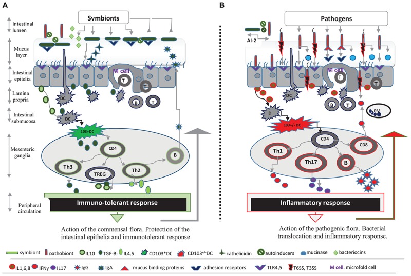
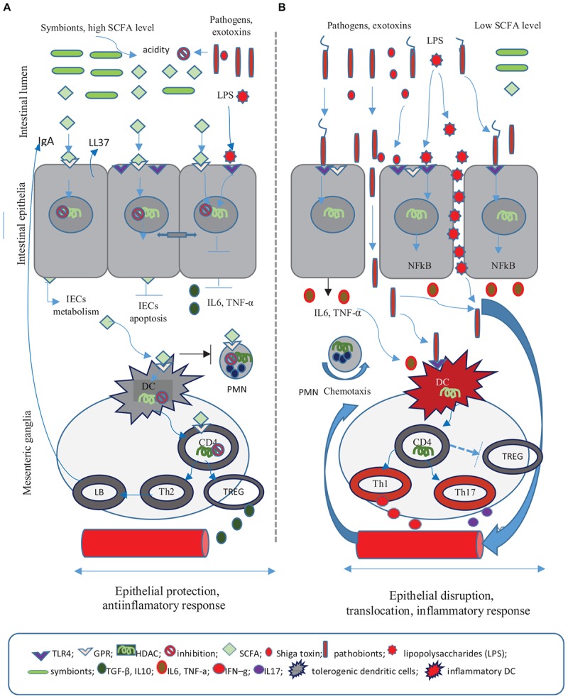
The intestinal microbiota is a complex microbial community, with diverse and stable populations hosted by the gastrointestinal tract since birth. This ecosystem holds multiple anti-infectious, anti-inflammatory, and immune modulating roles decisive for intestinal homeostasis. Among these, colonization resistance refers to the dynamic antagonistic interactions between commensals and pathogenic flora. Hence, gut bacteria compete for the same intestinal niches and substrates, while also releasing antimicrobial substances such as bacteriocines and changing the environmental conditions. Short chain fatty acids (SCFAs) generated in anaerobic conditions prompt epigenetic regulatory mechanisms that favor a tolerogenic immune response. In addition, the commensal flora is involved in the synthesis of bactericidal products, namely secondary biliary acids or antimicrobial peptides (AMPs) such as cathellicidin-LL37, an immunomodulatory, antimicrobial, and wound healing peptide. Gut microbiota is protected through symbiotic relations with the hosting organism and by quorum sensing, a specific cell-to-cell communication system. Any alterations of these relationships favor the uncontrollable multiplication of the resident pathobionts or external entero-pathogens, prompting systemic translocations, inflammatory reactions, or exacerbations of bacterial virulence mechanisms (T6SS, T3SS) and ultimately lead to gastrointestinal or systemic infections. The article describes the metabolic and immunological mechanisms through which the intestinal microbiota is both an ally of the organism against enteric pathogens and an enemy that favors the development of infections.
Keywords: bacterial enteropathogens; colonization resistance; commensal flora; infection; intestinal microbiota; mucosal immunity; short chain fatty acids.
Figures
Similar articles
-
The Roles of Inflammation, Nutrient Availability and the Commensal Microbiota in Enteric Pathogen Infection.Microbiol Spectr. 2015 Jun;3(3). doi: 10.1128/microbiolspec.MBP-0008-2014. Microbiol Spectr. 2015. PMID: 26185088
-
Infectious Threats, the Intestinal Barrier, and Its Trojan Horse: Dysbiosis.Front Microbiol. 2019 Aug 7;10:1676. doi: 10.3389/fmicb.2019.01676. eCollection 2019. Front Microbiol. 2019. PMID: 31447793 Free PMC article. Review.
-
Mechanism of the Gut Microbiota Colonization Resistance and Enteric Pathogen Infection.Front Cell Infect Microbiol. 2021 Dec 23;11:716299. doi: 10.3389/fcimb.2021.716299. eCollection 2021. Front Cell Infect Microbiol. 2021. PMID: 35004340 Free PMC article. Review.
-
The microbiota: a crucial mediator in gut homeostasis and colonization resistance.Front Microbiol. 2024 Aug 6;15:1417864. doi: 10.3389/fmicb.2024.1417864. eCollection 2024. Front Microbiol. 2024. PMID: 39165572 Free PMC article. Review.
-
Control of pathogens and pathobionts by the gut microbiota.Nat Immunol. 2013 Jul;14(7):685-90. doi: 10.1038/ni.2608. Nat Immunol. 2013. PMID: 23778796 Free PMC article. Review.
Cited by
-
Proteomic Characterization of the Cellular Effects of AhR Activation by Microbial Tryptophan Catabolites in Endotoxin-Activated Human Macrophages.Int J Environ Res Public Health. 2021 Sep 30;18(19):10336. doi: 10.3390/ijerph181910336. Int J Environ Res Public Health. 2021. PMID: 34639632 Free PMC article.
-
Mechanisms of medicinal, pharmaceutical, and immunomodulatory action of probiotics bacteria and their secondary metabolites against disease management: an overview.Folia Microbiol (Praha). 2024 Jun;69(3):549-565. doi: 10.1007/s12223-024-01155-2. Epub 2024 Mar 27. Folia Microbiol (Praha). 2024. PMID: 38532057 Review.
-
The Mycobiome of Bats in the American Southwest Is Structured by Geography, Bat Species, and Behavior.Microb Ecol. 2023 Oct;86(3):1565-1574. doi: 10.1007/s00248-023-02230-w. Epub 2023 May 1. Microb Ecol. 2023. PMID: 37126126
-
Luteolin Alleviates Ulcerative Colitis in Mice by Modulating Gut Microbiota and Plasma Metabolism.Nutrients. 2025 Jan 7;17(2):203. doi: 10.3390/nu17020203. Nutrients. 2025. PMID: 39861331 Free PMC article.
-
Alterations in bacterial metabolites, cytokines, and mucosal integrity in the caecum of broilers caused by feed additives and host-related factors.Front Physiol. 2022 Sep 12;13:935870. doi: 10.3389/fphys.2022.935870. eCollection 2022. Front Physiol. 2022. PMID: 36171972 Free PMC article.
References
Publication types
LinkOut - more resources
Full Text Sources
Other Literature Sources