

Coarctation of Aorta in Children
- PMID: 30761242
- PMCID: PMC6368362
- DOI: 10.7759/cureus.3690
Coarctation of Aorta in Children
Abstract
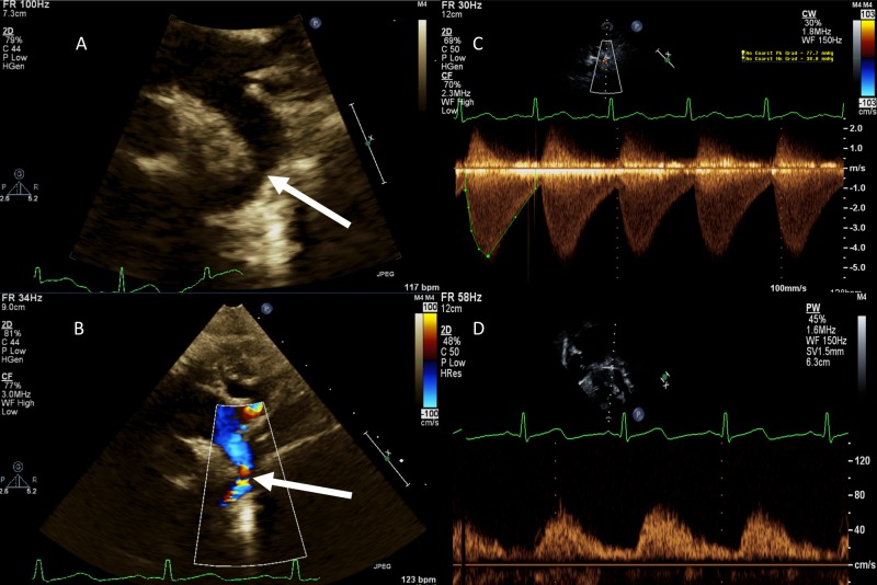
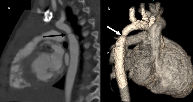
Coarctation of aorta (CoA) is a discrete narrowing in aorta causing obstruction to the flow of blood. It accounts for 6-8% of all congenital heart diseases. With advances in fetal echocardiography rate of prenatal diagnosis of coarctation of aorta has improved but it still remains a challenging diagnosis to make prenatally. Transthoracic echocardiography is mainstay of making initial diagnosis and routine follow-up. Cardiac magnetic resonance imaging (MRI) and computed tomography (CT) are great advanced imaging tools for two-dimensional and three-dimensional imaging of aortic arch in complex cases. Based on type of coarctation, size of patient, severity of lesion, and associated abnormalities various management options like surgical treatment, transcatheter balloon angioplasty and transcatheter stent implantation are available. There is significant improvement in long-term survival from pre-surgical era to post-surgical era. But, among the postsurgical era patients, the long-term survival has not significantly changed between older and contemporary cohort. Patients with coarctation of aorta need lifelong follow-up event after successful initial intervention.
Keywords: aortic coarctation; cardiac imaging; coarctation of aorta; pediatric coarctation.
Conflict of interest statement
The authors have declared that no competing interests exist.
Figures
Similar articles
-
Preoperative Physiology, Imaging, and Management of Coarctation of Aorta in Children.Semin Cardiothorac Vasc Anesth. 2019 Dec;23(4):379-386. doi: 10.1177/1089253219873004. Epub 2019 Sep 19. Semin Cardiothorac Vasc Anesth. 2019. PMID: 31535945 Review.
-
Management of adults with coarctation of aorta.World J Cardiol. 2020 May 26;12(5):167-191. doi: 10.4330/wjc.v12.i5.167. World J Cardiol. 2020. PMID: 32547712 Free PMC article. Review.
-
Diagnosis, imaging and clinical management of aortic coarctation.Heart. 2017 Aug;103(15):1148-1155. doi: 10.1136/heartjnl-2017-311173. Epub 2017 Apr 4. Heart. 2017. PMID: 28377475 Review.
-
Coarctation of the aorta.Curr Cardiol Rep. 2005 Nov;7(6):425-34. doi: 10.1007/s11886-005-0060-0. Curr Cardiol Rep. 2005. PMID: 16256011 Review.
-
Abnormalities of aortic arch shape, central aortic flow dynamics, and distensibility predispose to hypertension after successful repair of aortic coarctation.World J Pediatr Congenit Heart Surg. 2014 Oct;5(4):546-53. doi: 10.1177/2150135114551028. World J Pediatr Congenit Heart Surg. 2014. PMID: 25324252
Cited by
-
Aortic Arch Variants and Anomalies: Embryology, Imaging Findings, and Clinical Considerations.J Cardiovasc Imaging. 2022 Oct;30(4):231-262. doi: 10.4250/jcvi.2022.0058. J Cardiovasc Imaging. 2022. PMID: 36280266 Free PMC article. Review.
-
Coarctation of the Aorta and Late-Onset Sepsis With Supraventricular Tachycardia: A Clinical Case Review.Cureus. 2024 Jul 29;16(7):e65634. doi: 10.7759/cureus.65634. eCollection 2024 Jul. Cureus. 2024. PMID: 39205755 Free PMC article.
-
Simultaneous surgical treatment of aortic coarctation and aortic root disease: a report of case series.J Thorac Dis. 2023 Sep 28;15(9):5029-5036. doi: 10.21037/jtd-23-384. Epub 2023 Aug 30. J Thorac Dis. 2023. PMID: 37868868 Free PMC article.
-
Oxidative Stress and Endothelial Dysfunction: The Pathogenesis of Pediatric Hypertension.Int J Mol Sci. 2025 Jun 3;26(11):5355. doi: 10.3390/ijms26115355. Int J Mol Sci. 2025. PMID: 40508164 Free PMC article. Review.
-
320-slice Computed Tomography Angiography Imaging Findings and Follow-up in A Patient with Aortic Coarctation Misdiagnosed as Hypertension: A Case Report and Literature Review.Cureus. 2019 Dec 31;11(12):e6529. doi: 10.7759/cureus.6529. Cureus. 2019. PMID: 32010536 Free PMC article.
References
-
- Coarctation of aorta-management options and decision making. Doshi AR, Syamasundar Rao P. Pediat Therapeut. 2012;5:6.
-
- Clarence Crafoord: a giant in cardiothoracic surgery, the first to repair aortic coarctation. Kvitting JP, Olin CL. Ann Thorac Surg. 2009;87:342–346. - PubMed
-
- Coarctation of the aorta: strategies for improving outcomes. Nguyen L, Cook SC. Cardiol Clin. 2015;33:521–530. - PubMed
-
- Transluminal aortic balloon angioplasty for coarctation of the aorta in the newborn. Singer MI, Rowen M, Dorsey TJ. Am Heart J. 1982;103:131–132. - PubMed
Publication types
LinkOut - more resources
Full Text Sources