. 2019 Jan 8;9(2):291-303.
doi: 10.1002/2211-5463.12573. eCollection 2019 Feb.

Transferrin receptor 1 is required for enucleation of mouse erythroblasts during terminal differentiation

Affiliations

Transferrin receptor 1 is required for enucleation of mouse erythroblasts during terminal differentiation

Mamoru Aoto et al. FEBS Open Bio. .

Abstract

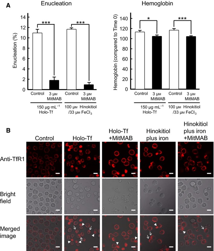
Enucleation is the process whereby the nucleus is extruded from the erythroblast during late stage mammalian erythropoiesis. However, the specific signaling pathways involved in this process remain unclear. To better understand the mechanisms underlying erythroblast enucleation, we investigated erythroblast enucleation using both the spleens of adult mice with phenylhydrazine-induced anemia and mouse fetal livers. Our results indicated that both iron-bound transferrin (holo-Tf) and the small-molecule iron transporter hinokitiol with iron ions (hinokitiol plus iron) promote hemoglobin synthesis and the enucleation of mouse spleen-derived erythroblasts. Although an antitransferrin receptor 1 (TfR1) monoclonal antibody inhibited both enucleation and hemoglobin synthesis promoted by holo-Tf, it inhibited only enucleation, but not hemoglobin synthesis, promoted by hinokitiol plus iron. Furthermore, siRNA against mouse TfR1 were found to suppress the enucleation of mouse fetal liver-derived erythroblasts, and the endocytosis inhibitor MitMAB inhibited enucleation, hemoglobin synthesis, and the internalization of TfR1 promoted by both types of stimuli. Collectively, our results suggest that TfR1, iron ions, and endocytosis play important roles in mouse erythroblast enucleation.

Keywords: enucleation; erythroblast; hinokitiol; iron; transferrin; transferrin receptor.


Figures

Figure 1
Figure 1
Holo‐Tf stimulates enucleation and hemoglobin synthesis in mouse spleen‐derived erythroblasts via TfR1. (A) Crude splenocytes and splenocytes after Percoll gradient centrifugation and subsequent CD45‐depletion were stained for PE‐anti‐Ter119, PE/Cy7‐anti‐CD71, and Pacific blue‐anti‐CD45 antibodies and analyzed by flow cytometry. Ter119‐positive cells in each fraction were further analyzed using FITC‐anti‐CD44 antibody. Purified CD45‐negative cells were used as mouse spleen‐derived erythroblast preparation. (B) Mouse spleen‐derived erythroblasts before or after incubation for 5 h were stained with PE‐anti‐Ter119 and SYTO16 and then analyzed by flow cytometry. (C) Enucleation and hemoglobin synthesis assays of mouse spleen‐derived erythroblasts in the presence of various concentrations of human holo‐Tf (one‐way ANOVA). Erythroblasts were incubated alone (open circles) or with 150 μg·mL−1 of human holo‐Tf (closed circles) for the indicated time (two‐way ANOVA). Purified mouse spleen‐derived erythroblasts were incubated for 5 h. Then, their hemoglobin content was quantified (Student's t‐test). (D) The total cell numbers (×106) and the number of reticulocytes after 5 h of culture in the presence/absence of holo‐Tf (Student's t‐test). The dashed lines show the starting cell number (3.0 × 10⁶) and the initial number of reticulocytes (1.3 × 10⁶), respectively. All experiments were repeated four times. (C, D) All data are the mean ± SEM. *P < 0.05, **P < 0.01, and ***p < 0.001 (compared to the control).
Figure 2
Figure 2
The anti‐TfR1 monoclonal antibody R17 208.2 blocks mouse erythroblast enucleation. (A) Enucleation assays of primary mouse spleen‐derived erythroblasts in the presence of 150 μg·mL−1 of human holo‐Tf and various concentrations of the R17 208.2 antibody or control IgM. (B) Hemoglobin assays of mouse spleen‐derived erythroblasts in the presence of 150 μg·mL−1 of human holo‐Tf and 6 μg·mL−1 of the R17 208.2 antibody or control IgM. (A, B) Experiments were repeated four times (one‐way ANOVA). All data are the mean ± SEM. *p < 0.05, **p < 0.01, and ***p < 0.001 (compared to the control). (C) Confirmation of mouse TfR1 or TfR2 expression on the cell surface of K562 cells by staining with FITC‐labeled human holo‐Tf. Analysis of the specificity of the R17 208.2 antibody. Parental K562, pcDNA3.1‐K562, mTfR1‐K562, and mTfR2‐K562 cells were stained with either the R17 208.2 antibody or control IgM, followed with the PE‐anti‐Rat IgM antibody. Then, cells were analyzed by flow cytometry. The R17 208.2 antibody recognized TfR1 but not TfR2.
Figure 3
Figure 3
Hinokitiol plus iron stimulates mouse spleen‐derived erythroblast enucleation. (A) Enucleation or hemoglobin assays of mouse spleen‐derived erythroblasts in the presence of 100 μm hinokitiol, 33 μm FeCl3, vehicle, or both 100 μm hinokitiol and 33 μm FeCl3. Experiments were performed eight or four times (one‐way ANOVA). (B) The total cell numbers (×106) and the number of reticulocytes after 5 h of culture in the presence/absence of hinokitiol plus iron. The dashed lines show the starting cell number (3.0 × 10⁶) and the initial number of reticulocytes (1.1 × 10⁶), respectively. Experiments were performed six times (one‐way ANOVA). (C) Hinokitiol plus iron did not negate the inhibitory effect of the R17 208.2 antibody (6 μg·mL−1) on 150 μg·mL−1 holo‐Tf‐promoted enucleation, but canceled the inhibitory effect on holo‐Tf‐promoted hemoglobin synthesis. Experiments were performed eight or 13 times (one‐way ANOVA). (D) The R17 208.2 antibody (6 μg·mL−1) inhibited 100 μm hinokitiol and 33 μm FeCl3‐promoted mouse spleen‐derived erythroblast enucleation, but not hemoglobin synthesis. Experiments were performed six times (one‐way ANOVA). (A–D) All data are the mean ± SEM. *p < 0.05 and **p < 0.01 (compared to the control).
Figure 4
Figure 4
The anti‐TfR1 monoclonal antibody R17 208.2 and siRNA for mouse TfR1 block mouse fetal liver‐derived erythroblast enucleation. (A) Results of enucleation assays of Ter119‐negative mouse fetal liver‐derived erythroblasts in medium containing 20% FBS and various concentrations of the R17 208.2 antibody or control IgM. Experiments were repeated four times (one‐way ANOVA). (B) Hinokitiol plus iron did not cancel the inhibitory effect of the R17 208.2 antibody (1.3 μg·mL−1) on mouse fetal liver‐derived erythroblast enucleation. Experiments were performed six times (one‐way ANOVA). (C) Mouse fetal liver‐derived erythroblasts were transfected with mouse TfR1 siRNA #1, #2, the GFP siRNA or negative control siRNA. Expression of mouse TfR1 protein or Ter119 antigen was assessed by flow cytometry with PE/Cy7‐anti‐TfR1 or PE‐anti‐Ter119. (D, E) Mouse fetal liver‐derived erythroblasts were transfected with mouse TfR1 siRNA #1, mouse TfR1 siRNA #2, the GFP siRNA or negative control siRNA, and then were cultured for 48 h. The ratio of enucleation was analyzed by flow cytometry. Experiments were repeated four times or six times (one‐way ANOVA). All data are the mean ± SEM. *p < 0.05, **p < 0.01, and ***p < 0.001 (compared to the control).
Figure 5
Figure 5
The endocytosis inhibitor MitMAB blocked enucleation, hemoglobin synthesis of mouse spleen‐derived erythroblasts, and the internalization of TfR1. (A) Enucleation or hemoglobin assays of mouse spleen‐derived erythroblasts in the absence or presence of 3 μm MitMAB. Experiments were performed six times (Student's t‐test). All data are the mean ± SEM. *p < 0.05, and ***p < 0.001 (compared to the control). (B) The endocytosis of TfR1 induced by holo‐Tf or hinokitiol plus iron was suppressed in the presence of dynamin inhibitor MitMAB. Mouse spleen‐derived erythroblasts were stimulated by holo‐Tf or hinokitiol plus iron in the presence or absence of 3 μm MitMAB, and 30 min later, the erythroblasts were immunostained with the anti‐TfR1 antibody followed by the RITC‐conjugated anti‐rabbit antibody (Top panel). The bright field images and the merged images are shown in middle panels and bottom panels, respectively. Arrows indicate the localization of TfR1 to the opposite side of the nucleus. Arrowheads indicate the localization of TfR1 to the cytoplasmic side immediately after enucleation. MitMAB suppressed the change of localization of TfR1 occurred after both stimulations. Scale bar, 10 μm.

Similar articles

Cited by

References

    1. Wu H, Liu X, Jaenisch R and Lodish HF (1995) Generation of committed erythroid BFU‐E and CFU‐E progenitors does not require erythropoietin or the erythropoietin receptor. Cell 83, 59–67. - PubMed
    1. Koury MJ and Bondurant MC (1988) Maintenance by erythropoietin of viability and maturation of murine erythroid precursor cells. J Cell Physiol 137, 65–74. - PubMed
    1. Ji P, Yeh V, Ramirez T, Murata‐Hori M and Lodish HF (2010) Histone deacetylase 2 is required for chromatin condensation and subsequent enucleation of cultured mouse fetal erythroblasts. Haematologica 95, 2013–2021. - PMC - PubMed
    1. Li X, Mei Y, Yan B, Vitriol E, Huang S, Ji P and Qiu Y (2017) Histone deacetylase 6 regulates cytokinesis and erythrocyte enucleation through deacetylation of formin protein mDia2. Haematologica 102, 984–994. - PMC - PubMed
    1. Ji P, Jayapal SR and Lodish HF (2008) Enucleation of cultured mouse fetal erythroblasts requires Rac GTPases and mDia2. Nat Cell Biol 10, 314–321. - PubMed

Publication types

MeSH terms