. 2019 Jan 19;9(2):348-363.
doi: 10.1002/2211-5463.12580. eCollection 2019 Feb.

Coenzyme Q10 protects against burn-induced mitochondrial dysfunction and impaired insulin signaling in mouse skeletal muscle

Affiliations

Coenzyme Q10 protects against burn-induced mitochondrial dysfunction and impaired insulin signaling in mouse skeletal muscle

Harumasa Nakazawa et al. FEBS Open Bio. .

Abstract

Mitochondrial dysfunction is associated with metabolic alterations in various disease states, including major trauma (e.g., burn injury). Metabolic derangements, including muscle insulin resistance and hyperlactatemia, are a clinically significant complication of major trauma. Coenzyme Q10 (CoQ10) is an essential cofactor for mitochondrial electron transport, and its reduced form acts as a lipophilic antioxidant. Here, we report that burn injury induces impaired muscle insulin signaling, hyperlactatemia, mitochondrial dysfunction (as indicated by suppressed mitochondrial oxygen consumption rates), morphological alterations of the mitochondria (e. g., enlargement, and loss of cristae structure), mitochondrial oxidative stress, and disruption of mitochondrial integrity (as reflected by increased mitochondrial DNA levels in the cytosol and circulation). All of these alterations were significantly alleviated by CoQ10 treatment compared with vehicle alone. These findings indicate that CoQ10 treatment is efficacious in protecting against mitochondrial dysfunction and insulin resistance in skeletal muscle of burned mice. Our data highlight CoQ10 as a potential new strategy to prevent mitochondrial damage and metabolic dysfunction in burn patients.

Keywords: burn injury; coenzyme Q10; insulin resistance; mitochondrial dysfunction; skeletal muscle.


Figures

Figure 1
Figure 1
Coenzyme Q10 mitigated burn‐induced decreases in mitochondrial oxygen consumption rate (OCR) and ATP content in skeletal muscle. ADP‐ and FCCP‐stimulated OCRs were significantly decreased by burn injury compared with sham‐burn, both of which were ameliorated by CoQ10 treatment (A–C). Similarly, complex I‐, complex II‐, and complex IV‐dependent OCRs were significantly decreased by burn injury compared with sham‐burn, all of which were ameliorated by CoQ10 (D–G). ATP content in skeletal muscle was suppressed by burn injury compared with sham‐burn, which was mitigated by CoQ10 (H). ADP, adenosine diphosphate; Oligo, oligomycin; FCCP, carbonyl cyanide 4‐(trifluoromethoxy) phenylhydrazone; Rote, rotenone; AA, antimycin A; Succ, succinate; Asc, ascorbate; TMPD, N,N,N9,N9‐tetramethyl‐p‐phenylenediamine. The data were compared with one‐way ANOVA followed by Tukey's multiple comparison test. All values are presented as mean ± SEM. *P < 0.05, **P < 0.01, ***P < 0.001. n = 4 mice per group (A‐G). n = 6 mice per group (H).
Figure 2
Figure 2
Coenzyme Q10 prevented burn injury‐induced morphological alterations of mitochondria in muse skeletal muscle. Burn injury induced the morphological alterations, enlargement (A–L) and loss of cristae structure (A–H, M, and N), in mitochondria, both of which were prevented by CoQ10. White arrows indicate loss of cristae structure (G). A, E: sham + vehicle; B, F: sham + CoQ10; C,G: burn + vehicle; D, H:burn + CoQ10. The data were compared with one‐way ANOVA followed by Tukey's multiple comparison test. All values are presented as mean ± SEM. **P < 0.01, ***P < 0.001. n = 3 mice per group.
Figure 3
Figure 3
Coenzyme Q10 prevented burn‐induced impaired mitochondrial respiratory supercomplex assembly in skeletal muscle. Respiratory supercomplexes in the mitochondria were separated by blue native PAGE followed by two‐dimensional SDS/PAGE and visualized using antibodies for NDUFA9 (a component of complex I) and UQCRC2 and RISP (components of complex III). The percentage of the complex I‐containing largest supercomplex (I/III 2/IV n) signal intensity was decreased in vehicle‐treated burned mice compared with sham‐burned mice (A). CoQ10 inhibited the burn‐induced alteration in the supercomplexes. On the other hand, total protein abundance of UQCRC2 was not altered by burn injury or FTI‐277 (B). SDHA (a component of complex II) was used as a control. IB: immunoblotting.
Figure 4
Figure 4
Effects of burn injury and CoQ10 on proteins involved in mitochondrial fusion in mouse skeletal muscle. The longest form of OPA1 (a) was induced by burn injury, which was ameliorated by CoQ10 (A, B). Protein expression of other isoforms (b–e) was not altered by burn injury or CoQ10 (A, C–E). MFN1, MFN2, OMA1, and YME1L1 protein expression was increased by burn injury (A, F–I). CoQ10 significantly decreased MFN1, MFN2, and OMA1 expression in burned mice. CoQ10 appears to decrease YME1L1 expression in burned mice, but there was no statistical difference. The data were compared with one‐way ANOVA followed by Tukey's multiple comparison test. All values are presented as mean ± SEM. **P < 0.01, ***P < 0.001, NS: not significant. n = 6 mice per group.
Figure 5
Figure 5
Effects of burn injury and CoQ10 on proteins involved in mitochondrial fission in mouse skeletal muscle. Burn injury increased phosphorylated Drp1 (p‐Drp1), which was inhibited by CoQ10 (A, B). Burn injury increased Drp1 protein expression compared with sham‐burn (A, C). When treated with CoQ10, Drp1 expression was no longer increased relative to sham‐burned mice. Neither burn injury nor CoQ10 altered Fis1 expression (A, D). Burn injury increased Parkin and PINK1 protein expression, which was ameliorated by CoQ10 (A, E, and F). The data were compared with one‐way ANOVA followed by Tukey's multiple comparison test. All values are presented as mean ± SEM. *P < 0.05, ***P < 0.001, NS, not significant. n = 6 mice per group.
Figure 6
Figure 6
Coenzyme Q10 attenuated burn injury‐induced mitochondrial oxidative stress and mitochondrial unfolded protein response (mtUPR) in mouse skeletal muscle. To assess mitochondrial oxidative stress, oxidized (carbonylated) proteins in the mitochondria were measured. Burn injury increased oxidized proteins in the mitochondria, which was prevented by CoQ10 (A and B). Oxidized (carbonylated) proteins were detected only when incubated with DNPH, confirming the specificity. To assess mtUPR, protein expression of HSP90, HSP60, GRP75, and CLPP was measured. These protein expressions in skeletal muscle were significantly increased by burn injury, which were ameliorated by CoQ10 (C–F, H). LONP1 expression was not altered by burn and CoQ10 (C and G). DNPH: 2,4‐dinitrophenylhydrazine. The data were compared with one‐way ANOVA followed by Tukey's multiple comparison test. All values are presented as mean ± SEM. *P < 0.05, **P < 0.01, ***P < 0.001. n = 6 mice per group.
Figure 7
Figure 7
Coenzyme Q10 inhibited the effects of burn injury on mtDNA levels and circulating HMGB1. Burn injury decreased mtDNA‐to‐nDNA ratio, an indicator of total mtDNA content, compared with sham‐burn, which was mitigated by CoQ10 (A). In contrast, burn injury increased mtDNA levels in the cytosol (B) and plasma (C), both of which were ameliorated by CoQ10. Similar to plasma mtDNA, plasma HMGB1 concentration was increased by burn injury, which was inhibited by CoQ10 (D). The data were compared with one‐way ANOVA followed by Tukey's multiple comparison test. All values are presented as mean ± SEM. *P < 0.05, **P < 0.01, ***P < 0.001. n = 6 mice per group.
Figure 8
Figure 8
Coenzyme Q10 inhibited burn injury‐induced NLRP3 inflammasome activation in skeletal muscle. Burn increased protein expression of ASC and NLRP3, components of NLRP3 inflammasome (A–C). CoQ10 inhibited burn‐induced expression of ASC and NLRP3. Burn injury increased expression of pro‐caspase‐1 and cleaved caspase‐1 (p10 and p20) compared with sham‐burn (D–G). CoQ10 did not significantly decrease pro‐caspase‐1 expression, but inhibited burn injury‐induced expression of cleaved caspase‐1 (p10 and p20). Similarly, burn injury increased cleaved IL‐1β, which was ameliorated by CoQ10 (H–J). The data were compared with one‐way ANOVA followed by Tukey's multiple comparison test. All values are presented as mean ± SEM. *P < 0.05, **P < 0.01, ***P < 0.001. n = 6 mice per group.
Figure 9
Figure 9
Coenzyme Q10 inhibited burn injury‐induced proinflammatory expression in skeletal muscle. Burn injury increased the mRNA levels of IL‐1α (A), IL‐1β (B), IFN‐γ (C), TNF‐α (D), TLR4 (E), and caspase‐11 (F), which were inhibited by CoQ10. The data were compared with one‐way ANOVA followed by Tukey's multiple comparison test. All values are presented as mean ± SEM. *P < 0.05, **P < 0.01, ***P < 0.001. n = 6 mice per group.
Figure 10
Figure 10
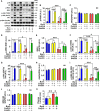
Coenzyme Q10 prevented burn injury‐induced impaired insulin signaling and hyperlactatemia in mouse skeletal muscle. Burn injury inhibited insulin‐stimulated phosphorylation of IR (B), IRS1 (D), Akt (F and G), and GSK‐3β (I) at 3 days after burn injury compared with sham‐burn. CoQ10 significantly ameliorated insulin‐stimulated phosphorylation of IR (B), IRS1 (D), Akt (F and G), and GSK‐3β (I) in burned mice. Protein expression of IRS1 was suppressed by burn injury, and CoQ10 increased IRS1 expression in burned mice (E). On the other hand, protein expression of IR (C), Akt (H), and GSK‐3β (J) was not altered by burn injury or CoQ10. Plasma lactate level was significantly increased at 3 days after burn injury, and CoQ10 supplementation ameliorated burn‐induced hyperlactatemia (K). The data were compared with two‐way ANOVA followed by Tukey's multiple comparison test. All values are presented as mean ± SEM. *P < 0.05, **P < 0.01, ***P < 0.001, NS: not significant. n = 4 mice per group (A–J), n = 6 mice per group (K).

Similar articles

Cited by

References

    1. Chondronikola M, Meyer WJ, Sidossis LS, Ojeda S, Huddleston J, Stevens P, Borsheim E, Suman OE, Finnerty CC and Herndon DN (2014) Predictors of insulin resistance in pediatric burn injury survivors 24 to 36 months postburn. J Burn Care Res 35, 409–415. - PMC - PubMed
    1. Cochran A, Edelman LS, Saffle JR and Morris SE (2007) The relationship of serum lactate and base deficit in burn patients to mortality. J Burn Care Res 28, 231–240. - PubMed
    1. Gore DC, Ferrando A, Barnett J, Wolf SE, Desai M, Herndon DN, Goodwin C & Wolfe RR (2000) Influence of glucose kinetics on plasma lactate concentration and energy expenditure in severely burned patients. J Trauma 49, 673–677; discussion 677‐8. - PubMed
    1. Merritt EK, Thalacker‐Mercer A, Cross JM, Windham ST, Thomas SJ and Bamman MM (2013) Increased expression of atrogenes and TWEAK family members after severe burn injury in nonburned human skeletal muscle. J Burn Care Res 34, e297–e304. - PMC - PubMed
    1. Brealey D, Brand M, Hargreaves I, Heales S, Land J, Smolenski R, Davies NA, Cooper CE and Singer M (2002) Association between mitochondrial dysfunction and severity and outcome of septic shock. Lancet 360, 219–223. - PubMed

Publication types