. 2019 Jan 29:6:6.
doi: 10.3389/fmed.2019.00006. eCollection 2019.

Intracellular Streptococcal Uptake and Persistence: A Potential Cause of Erysipelas Recurrence

Affiliations

Intracellular Streptococcal Uptake and Persistence: A Potential Cause of Erysipelas Recurrence

Fatma Jendoubi et al. Front Med (Lausanne). .

Abstract

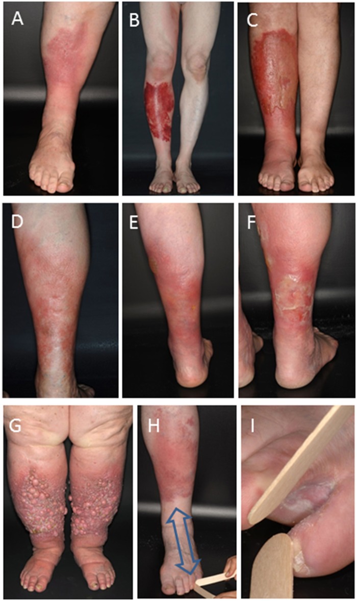
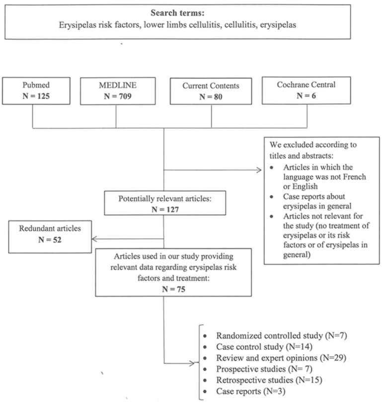
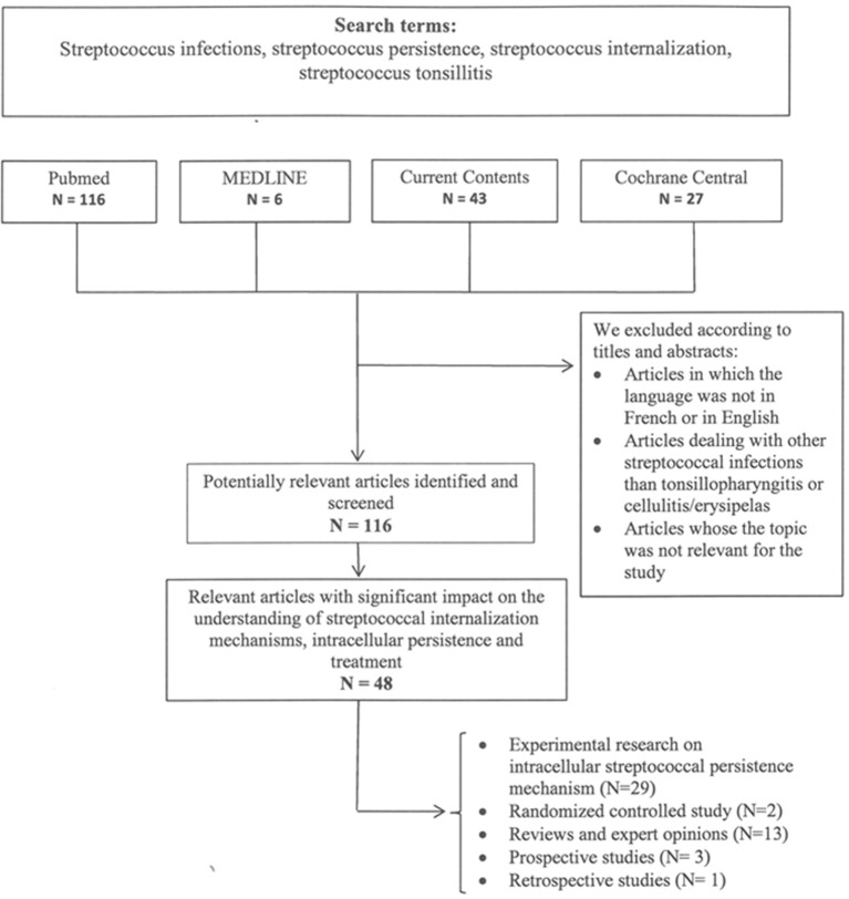
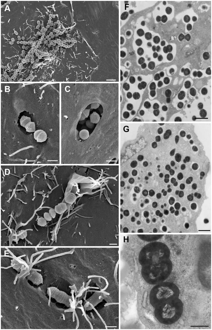
Erysipelas is a severe streptococcal infection of the skin primarily spreading through the lymphatic vessels. Penicillin is the treatment of choice. The most common complication consists in relapses which occur in up to 40% or more of patients despite appropriate antibiotic treatment. They cause lymphatic damage resulting in irreversible lymphedema and ultimately elephantiasis nostras and lead to major health restrictions and high socio-medical costs. Prevention of relapses is an unmet need, because even long-term prophylactic penicillin application does eventually not reduce the risk of recurrence. In this article we assess risk factors and causes of erysipelas recurrence. A systematic literature search for clinical studies addressing potential causes and measures for prevention of erysipelas recurrence was combined with a review of experimental and clinical data assessing the ability and clinical relevance of streptococci for intracellular uptake and persistence. The literature review found that venous insufficiency, lymphedema, and intertrigo from fungal infections are considered to be major risk factors for recurrence of erysipelas but cannot adequately explain the high recurrence rate. As hitherto unrecognized likely cause of erysipelas relapses we identify the ability of streptococci for intracellular uptake into and persistence within epithelial and endothelial cells and macrophages. This creates intracellular streptococcal reservoirs out of reach of penicillins which do not reach sufficient bactericidal intracellular concentrations. Incomplete streptococcal elimination due to intracellular streptococcal persistence has been observed in various deep tissue infections and is considered as cause of relapsing streptococcal pharyngitis despite proper antibiotic treatment. It may also serves as endogenous infectious source of erysipelas relapses. We conclude that the current antibiotic treatment strategies and elimination of conventional risk factors employed in erysipelas management are insufficient to prevent erysipelas recurrence. The reactivation of streptococcal infection from intracellular reservoirs represents a plausible explanation for the frequent occurrence erysipelas relapses. Prevention of erysipelas relapses therefore demands for novel antibiotic strategies capable of eradicating intracellular streptococcal persistence.

Keywords: cellulitis; erysipelas; intracellular streptococcal persistence; intracellular streptococcal uptake; penicillin prophylaxis; relapse; risk factors; treatment.


Figures

Figure 1
Figure 1
Clinical manifestation of erysipelas and erysipelas relapses. Typical inflammatory (A) or hemorrhagic (B) erysipelas of the lower leg. (C) Increase in lymphedema following several relapses in bullous erysipelas as indicated by the circumferential increase of the left ankle and non-pitting edema of the forefoot. (D) First erysipelas episode and two relapses two (E) and three (F) years after the first episode typically occurring in the same site of a female patient. (G) Late-stage excessive lymphedema with thickened fold of skin at the base of the second toe that cannot be lifted (positive Stemmer sign test), dermal fibrosis, and formation of numerous fibrotic nodes from multiple episodes of relapsing erysipelas. (H,I) Toe-web inoculation site of a first erysipelas episode of the left lower leg. Note the typical centripetal distance (arrow) between bacterial inoculation site and erysipelas.
Figure 2
Figure 2
Search terms and flow diagram of literature analysis for recurrence of erysipelas. N designates the number of articles.
Figure 3
Figure 3
Search terms and flow diagram of literature analysis for intracellular streptococcal uptake and persistence. N designates the number of articles.
Figure 4
Figure 4
The intracellular life-style of Group A streptococci. (A) Adhesion to host cell membrane of HEp-2 cell via adhesins. (B,C) Invasion into HEp-2 cells via invasins by co-opting caveolae for forming large invaginations for SfbI-carrying streptococci. (D) Invasion through signaling events which trigger cytoskeletal rearrangements like membrane ruffling. (E) Adherent streptococci are overgrown by host cell microvilli which fuse to form membrane flaps that subsequently cover streptococci. (F) Streptococci residing intracellularly exclusively in membrane-bound phagosome-like compartments after short infection time (cell culture model). (G) Release of streptococci out of the membrane-surrounded compartment into the host cell cytoplasm after longer infection time where they replicate and persist (cell culture model). (H) In vivo mouse model providing evidence of streptococci residing and replicating free in the cytoplasm and supporting the observations generated in cell culture. Bars represent 2 μm in (A) 1 μm in (F), and G, 0.5 μm in (B–E,H). All images were taken from infection experiments performed in cell culture models and in a mouse model at the Helmholtz Centre for Infection Research. Experimental procedures were previously described (81, 82). (A–E) represent field emission scanning electron microscopic images; Figures (F–H) are transmission electron microscopic images of ultrathin sections of resin embedded samples.

Similar articles

Cited by

References

    1. Bernard P, Bedane C, Mounier M, Denis F, Catanzano G, Bonnetblanc JM. Streptococcal cause of erysipelas and cellulitis in adults. A microbiologic study using a direct immunofluorescence technique. Arch Dermatol. (1989) 125:779–82. 10.1001/archderm.1989.01670180051004 - DOI - PubMed
    1. Eriksson B, Jorup-Ronstrom C, Karkkonen K, Sjoblom AC, Holm SE. Erysipelas: clinical and bacteriologic spectrum and serological aspects. Clin Infect Dis. (1996) 23:1091–8. 10.1093/clinids/23.5.1091 - DOI - PubMed
    1. Siljander T, Karppelin M, Vahakuopus S, Syrjanen J, Toropainen M, Kere J, et al. . Acute bacterial, nonnecrotizing cellulitis in Finland: microbiological findings. Clin Infect Dis. (2008) 46:855–61. 10.1086/527388 - DOI - PubMed
    1. Jeng A, Beheshti M, Li J, Nathan R. The role of beta-hemolytic streptococci in causing diffuse, nonculturable cellulitis: a prospective investigation. Medicine (2010) 89:217–26. 10.1097/MD.0b013e3181e8d635 - DOI - PubMed
    1. Stevens DL, Bryant AE. Impetigo, erysipelas and cellulitis. In: Ferretti JJ, Stevens DL, Fischetti VA, editors. Streptococcus pyogenes: Basic Biology to Clinical Manifestations. Oklahoma City, OK: University of Oklahoma Health Sciences Center; (2016). p. 723–40. - PubMed