Review
. 2019 Aug;208(3-4):431-438.
doi: 10.1007/s00430-019-00581-1. Epub 2019 Feb 14.

HCMV latency: what regulates the regulators?

Affiliations
Review

HCMV latency: what regulates the regulators?

Elizabeth Elder et al. Med Microbiol Immunol. 2019 Aug.

Abstract

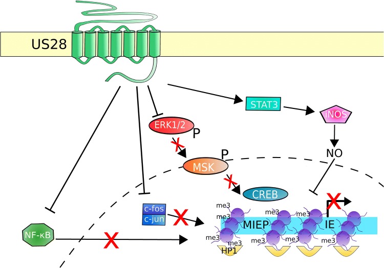
Human cytomegalovirus (HCMV) latency and reactivation is regulated by the chromatin structure at the major immediate early promoter (MIEP) within myeloid cells. Both cellular and viral factors are known to control this promoter during latency, here we will review the known mechanisms for MIEP regulation during latency. We will then focus on the virally encoded G-protein coupled receptor, US28, which suppresses the MIEP in early myeloid lineage cells. The importance of this function is underlined by the fact that US28 is essential for HCMV latency in CD34+ progenitor cells and CD14+ monocytes. We will describe cellular signalling pathways modulated by US28 to direct MIEP suppression during latency and demonstrate how US28 is able to 'regulate the regulators' of HCMV latency. Finally, we will describe how cell-surface US28 can be a target for antiviral therapies directed at the latent viral reservoir.

Keywords: Cell signalling; Chromatin; Cytomegalovirus; Latency; US28; Viral reservoir.


Conflict of interest statement

The authors declare they have no conflict of interest.

Figures

Fig. 1
Fig. 1
Regulation of HCMV latency and reactivation during myeloid differentiation. HCMV infects CD34+ progenitor cells and establishes latency (top left). The HCMV genome is maintained in the nucleus as an episome (blue circle) and is chromatinised. The MIEP (represented bottom left) is prevented from driving IE gene expression by a repressive chromatin state. Histones (purple) are trimethylated (me3) at H3K9 and H3K27. The repressive factor HP1 associates with the MIEP, as do ERF and YY1, and KAP1 acts to suppress the MIEP from distal binding sites. Latency-associated viral factors (listed) contribute to MIEP suppression, and the activatory factor pp71 is excluded from the nucleus. During differentiation-induced reactivation in mature dendritic cells or macrophages (top right), transcription of IE genes is activated leading to full lytic replication and release of infectious virions. As a result of differentiation, the chromatin structure around the MIEP is more open (bottom right), and activatory histone marks including histone acetylation (Ac) and H3-serine-10-phosphorylation (S10P) are present. Activated CREB and NF-κB become associated with the MIEP, as do histone acetyl transferases (HATs). Several viral factors are reported to be important for reactivation in myeloid cells, including LUNA, UL7, and certain members of the ULb’ family
Fig. 2
Fig. 2
US28 controls several signaling pathways to suppress the MIEP in early myeloid lineage cells. US28 is present at the cell surface, and probably other membranes, of latently infected cells. Here, it attenuates several signaling pathways and transcription factors, including NF-κB, c-fos, and ERK1/2. NF-κB can no longer enter the nucleus (dashed line), nor bind and activate the MIEP. c-fos typically forms a dimer with c-jun to form the AP1 complex; US28 causes loss of c-fos, the AP1 complex does not form and thus cannot activate the MIEP. Attenuation of ERK1/2 causes loss of ERK1/2 phosphorylation (P) and subsequent activation of MSK and, therefore, MSK does not phosphorylate and activate CREB. Inactive CREB cannot activate the MIEP. US28 is also reported to activate the STAT3-iNOS signaling axis, leading to nitric oxide (NO) production. NO suppresses the MIEP in myeloid cells by unknown mechanisms. By these, and probably other pathways, US28 helps establish and maintain a repressive chromatin structure at the MIEP, and a lack of IE gene expression

References

    1. Taylor-Wiedeman J, Sissons JGP, Borysiewicz LK, Sinclair JH. Monocytes are a major site of persistence of human cytomegalovirus in peripheral blood mononuclear cells. J Gen Virol. 1991;72:2059–2064. doi: 10.1099/0022-1317-72-9-2059. - DOI - PubMed
    1. Mendelson M, Monard S, Sissons P, Sinclair J. Detection of endogenous human cytomegalovirus in CD34 + bone marrow progenitors. J Gen Virol. 1996;77:3099–3102. doi: 10.1099/0022-1317-77-12-3099. - DOI - PubMed
    1. Kondo K, Xu J, Mocarski ES. Human cytomegalovirus latent gene expression in granulocyte-macrophage progenitors in culture and in seropositive individuals. Proc Natl Acad Sci USA. 1996;93:11137–11142. doi: 10.1073/pnas.93.20.11137. - DOI - PMC - PubMed
    1. Hahn G, Jores R, Mocarski ES. Cytomegalovirus remains latent in a common precursor of dendritic and myeloid cells. Proc Natl Acad Sci USA. 1998;95:3937–3942. doi: 10.1073/pnas.95.7.3937. - DOI - PMC - PubMed
    1. Reeves MB, Sinclair JH. Circulating dendritic cells isolated from healthy seropositive donors are sites of human cytomegalovirus reactivation in vivo. J Virol. 2013;87:10660–10667. doi: 10.1128/JVI.01539-13. - DOI - PMC - PubMed

LinkOut - more resources