

Phosphodiesterase Inhibitors Revert Axonal Dystrophy in Friedreich's Ataxia Mouse Model
- PMID: 30761510
- PMCID: PMC6554462
- DOI: 10.1007/s13311-018-00706-z
Phosphodiesterase Inhibitors Revert Axonal Dystrophy in Friedreich's Ataxia Mouse Model
Abstract
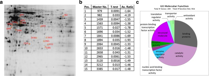
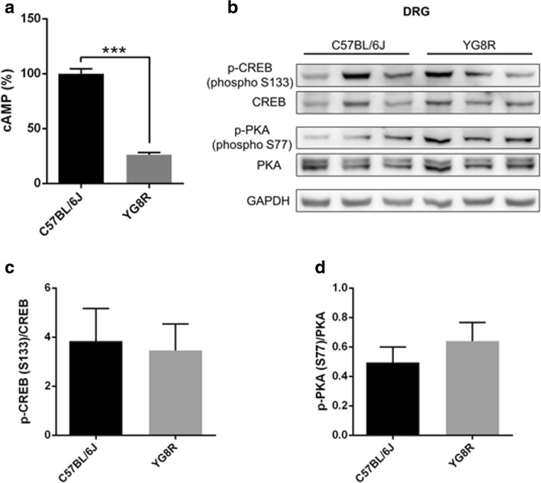
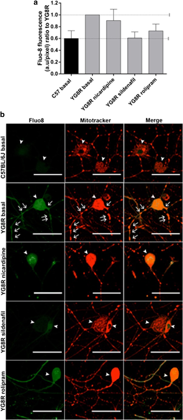
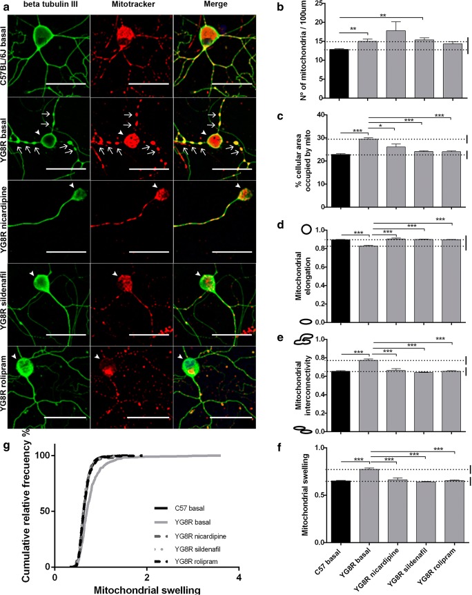
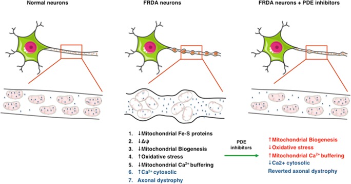
Friedreich's ataxia (FRDA) is a neurodegenerative disorder caused by an unstable GAA repeat expansion within intron 1 of the FXN gene and characterized by peripheral neuropathy. A major feature of FRDA is frataxin deficiency with the loss of large sensory neurons of the dorsal root ganglia (DRG), namely proprioceptive neurons, undergoing dying-back neurodegeneration with progression to posterior columns of the spinal cord and cerebellar ataxia. We used isolated DRGs from a YG8R FRDA mouse model and C57BL/6J control mice for a proteomic study and a primary culture of sensory neurons from DRG to test novel pharmacological strategies. We found a decreased expression of electron transport chain (ETC) proteins, the oxidative phosphorylation (OXPHOS) system and antioxidant enzymes, confirming a clear impairment in mitochondrial function and an oxidative stress-prone phenotype. The proteomic profile also showed a decreased expression in Ca2+ signaling related proteins and G protein-coupled receptors (GPCRs). These receptors modulate intracellular cAMP/cGMP and Ca2+ levels. Treatment of frataxin-deficient sensory neurons with phosphodiesterase (PDE) inhibitors was able to restore improper cytosolic Ca2+ levels and revert the axonal dystrophy found in DRG neurons of YG8R mice. In conclusion, the present study shows the effectiveness of PDE inhibitors against axonal degeneration of sensory neurons in YG8R mice. Our findings indicate that PDE inhibitors may become a future FRDA pharmacological treatment.
Keywords: Ca2+ signaling; FRDA; G protein-coupled receptor (GPCR); PDE inhibitors; axonal degeneration.
Conflict of interest statement
The authors declare that they have no competing interests.
Figures
Similar articles
-
Two different pathogenic mechanisms, dying-back axonal neuropathy and pancreatic senescence, are present in the YG8R mouse model of Friedreich's ataxia.Dis Model Mech. 2016 Jun 1;9(6):647-57. doi: 10.1242/dmm.024273. Epub 2016 Apr 14. Dis Model Mech. 2016. PMID: 27079523 Free PMC article.
-
Frataxin gene editing rescues Friedreich's ataxia pathology in dorsal root ganglia organoid-derived sensory neurons.Nat Commun. 2020 Aug 21;11(1):4178. doi: 10.1038/s41467-020-17954-3. Nat Commun. 2020. PMID: 32826895 Free PMC article.
-
Transplantation of wild-type mouse hematopoietic stem and progenitor cells ameliorates deficits in a mouse model of Friedreich's ataxia.Sci Transl Med. 2017 Oct 25;9(413):eaaj2347. doi: 10.1126/scitranslmed.aaj2347. Sci Transl Med. 2017. PMID: 29070698 Free PMC article.
-
Friedreich's ataxia: pathology, pathogenesis, and molecular genetics.J Neurol Sci. 2011 Apr 15;303(1-2):1-12. doi: 10.1016/j.jns.2011.01.010. J Neurol Sci. 2011. PMID: 21315377 Free PMC article. Review.
-
Mitochondrial pathophysiology in Friedreich's ataxia.J Neurochem. 2013 Aug;126 Suppl 1:53-64. doi: 10.1111/jnc.12303. J Neurochem. 2013. PMID: 23859341 Review.
Cited by
-
A new FRDA mouse model [Fxn null:YG8s(GAA) > 800] with more than 800 GAA repeats.Front Neurosci. 2023 Jan 26;17:930422. doi: 10.3389/fnins.2023.930422. eCollection 2023. Front Neurosci. 2023. PMID: 36777637 Free PMC article.
-
Cofilin dysregulation alters actin turnover in frataxin-deficient neurons.Sci Rep. 2020 Mar 23;10(1):5207. doi: 10.1038/s41598-020-62050-7. Sci Rep. 2020. PMID: 32251310 Free PMC article.
-
Molecular Mechanisms and Therapeutics for the GAA·TTC Expansion Disease Friedreich Ataxia.Neurotherapeutics. 2019 Oct;16(4):1032-1049. doi: 10.1007/s13311-019-00764-x. Neurotherapeutics. 2019. PMID: 31317428 Free PMC article. Review.
-
Alpha-lipoic acid supplementation improves pathological alterations in cellular models of Friedreich ataxia.Orphanet J Rare Dis. 2025 Aug 23;20(1):453. doi: 10.1186/s13023-025-03990-z. Orphanet J Rare Dis. 2025. PMID: 40849475
-
BRUCE silencing leads to axonal dystrophy by repressing autophagosome-lysosome fusion in Alzheimer's disease.Transl Psychiatry. 2021 Aug 5;11(1):421. doi: 10.1038/s41398-021-01427-2. Transl Psychiatry. 2021. PMID: 34354038 Free PMC article.
References
-
- Campuzano V, Montermini L, Molto MD, Pianese L, Cossee M, Cavalcanti F et al. Friedreich’s ataxia: autosomal recessive disease caused by an intronic GAA triplet repeat expansion. Science (New York NY). 1996;271(5254):1423–7. - PubMed
Publication types
MeSH terms
Substances
Grants and funding
- PI11/00678/Instituto de Salud Carlos III/International
- SAF2015-66625-R/Secretar?a de Estado de Investigaci?n, Desarrollo e Innovaci?n/International
- PROMETEOII/2014/067/Generalitat Valenciana/International
- PROMETEOII/2014/029/Generalitat Valenciana/International
- ACIF/2014/090/Generalitat Valenciana/International
LinkOut - more resources
Full Text Sources
Other Literature Sources
Medical
Miscellaneous
