. 2019 Jul;67(7):1277-1295.
doi: 10.1002/glia.23602. Epub 2019 Feb 13.

Independent and cooperative roles of the Mek/ERK1/2-MAPK and PI3K/Akt/mTOR pathways during developmental myelination and in adulthood

Affiliations

Independent and cooperative roles of the Mek/ERK1/2-MAPK and PI3K/Akt/mTOR pathways during developmental myelination and in adulthood

Akihiro Ishii et al. Glia. 2019 Jul.

Abstract

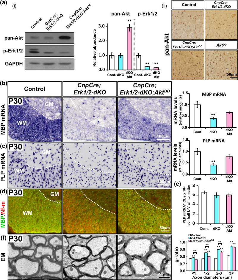
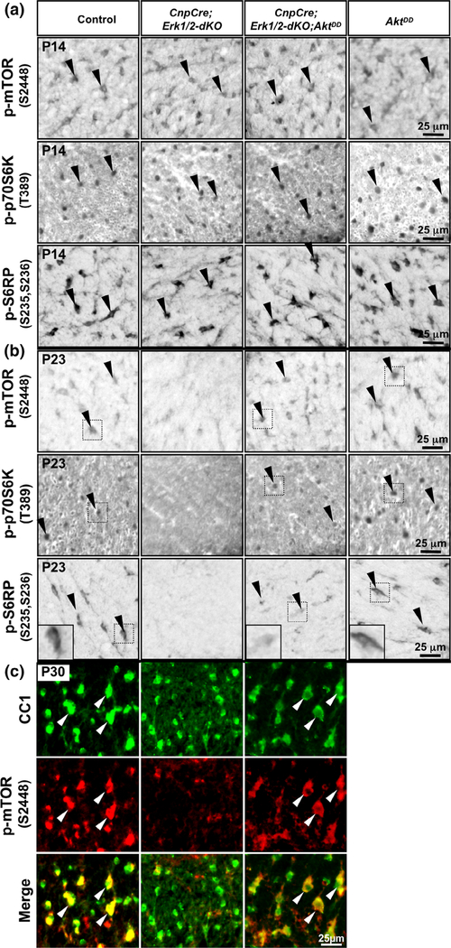
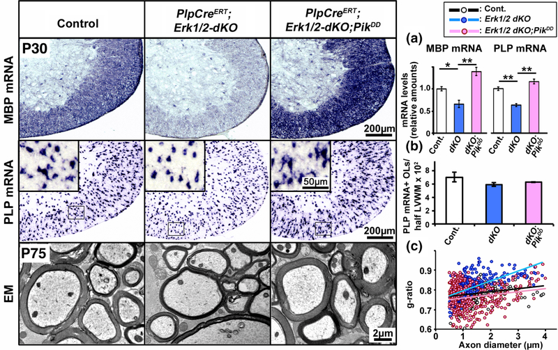
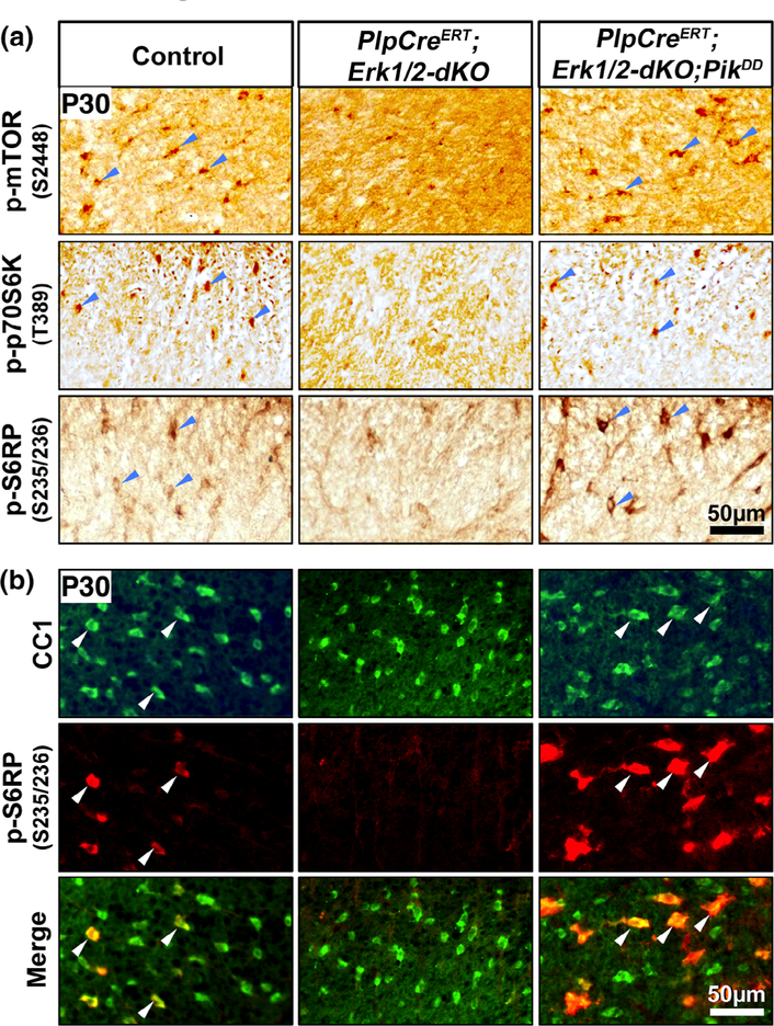
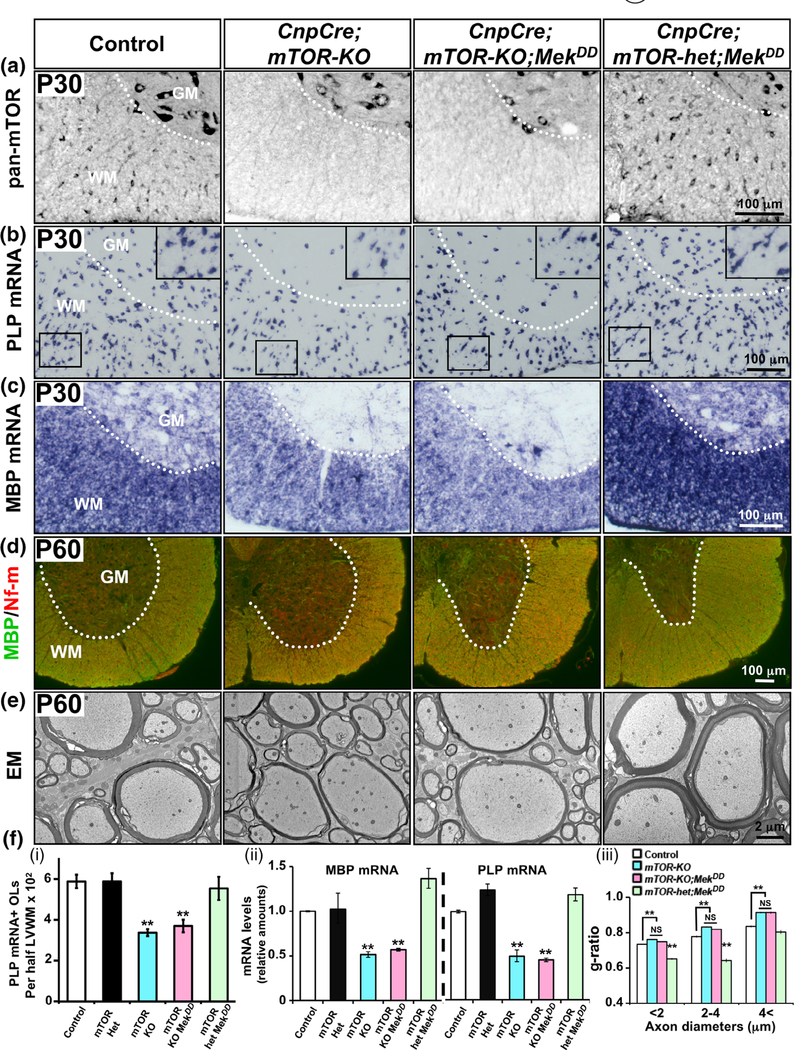
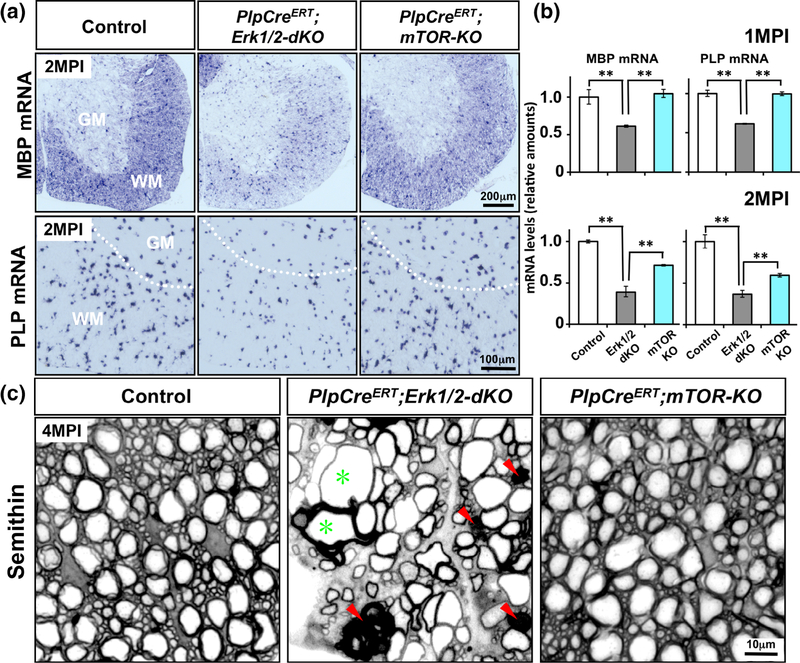
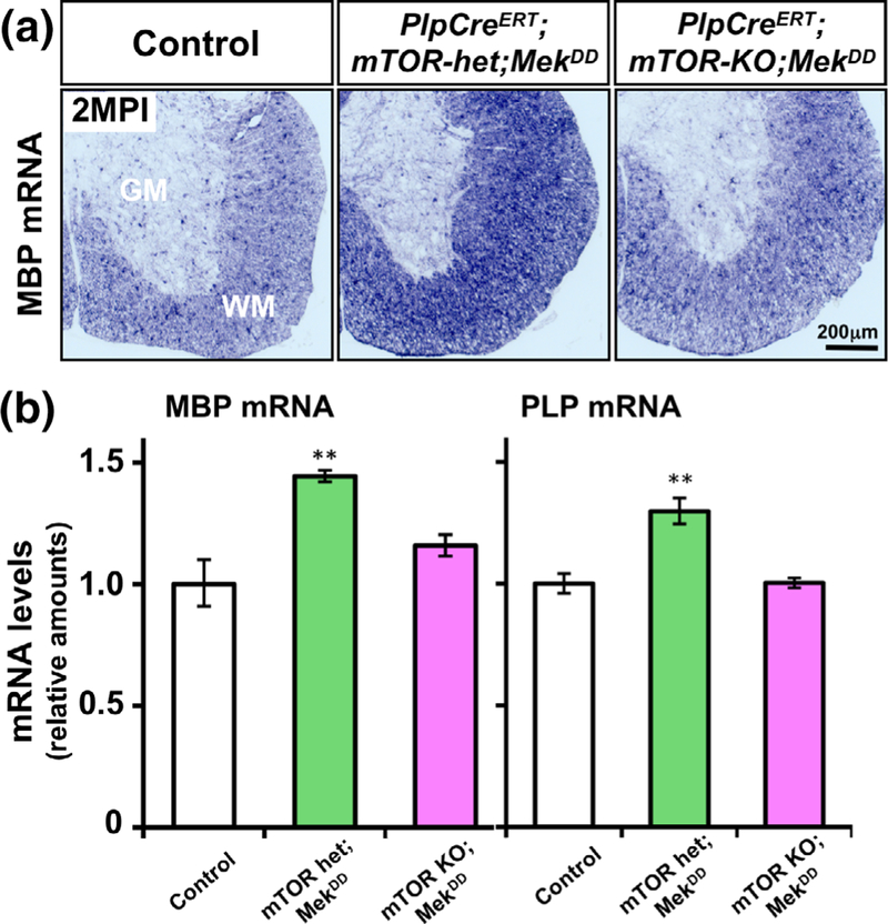
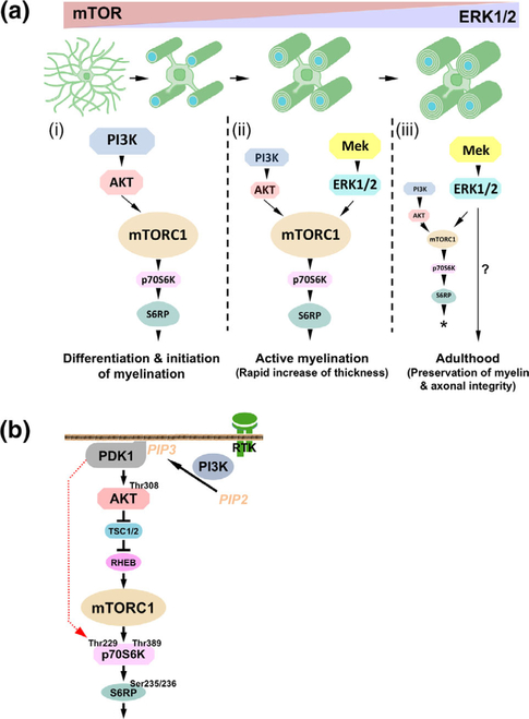
Multiple extracellular and intracellular signals regulate the functions of oligodendrocytes as they progress through the complex process of developmental myelination and then maintain a functionally intact myelin sheath throughout adult life, preserving the integrity of the axons. Recent studies suggest that Mek/ERK1/2-MAPK and PI3K/Akt/mTOR intracellular signaling pathways play important, often overlapping roles in the regulation of myelination. However, it remains poorly understood whether they function independently, sequentially, or converge using a common mechanism to facilitate oligodendrocyte differentiation, myelin growth, and maintenance. To address these questions, we analyzed multiple genetically modified mice and asked whether the deficits due to the conditional loss-of-function of ERK1/2 or mTOR could be abrogated by simultaneous constitutive activation of PI3K/Akt or Mek, respectively. From these studies, we concluded that while PI3K/Akt, not Mek/ERK1/2, plays a key role in promoting oligodendrocyte differentiation and timely initiation of myelination through mTORC1 signaling, Mek/ERK1/2-MAPK functions largely independently of mTORC1 to preserve the integrity of the myelinated axons during adulthood. However, to promote the efficient growth of the myelin sheath, these two pathways cooperate with each other converging at the level of mTORC1, both in the context of normal developmental myelination or following forced reactivation of the myelination program during adulthood. Thus, Mek/ERK1/2-MAPK and the PI3K/Akt/mTOR signaling pathways work both independently and cooperatively to maintain a finely tuned, temporally regulated balance as oligodendrocytes progress through different phases of developmental myelination into adulthood. Therapeutic strategies aimed at targeting remyelination in demyelinating diseases are expected to benefit from these findings.

Keywords: differentiation; myelin; myelination; myelinogenesis; oligodendrocyte.


Figures

FIGURE 1
FIGURE 1
Sustained overactivation of Akt in oligodendrocytes partially abrogated the deficit in myelin gene expression and myelin growth in mice lacking ERK1/2. (a (i)) Immuno-blot analysis of equal amounts of total proteins from homogenates of spinal cord white matter and quantification of the band intensity show that pan-Akt levels are similar in control (Cont.) and CnpCre;Erk1/2-dKO (dKO), but significantly increased in the CnpCre;Erk1/2-dKO;AktDD (dKOAkt) mice. p-Erk1/2 levels are significantly reduced in both CnpCre;Erk1/2-dKO and CnpCre; Erk1/2-dKO;AktDD compared to control. GAPDH, used as a loading control, does not show a change. (a (ii)) Immunolabeling of spinal cord sections for pan-Akt shows that compared to control and CnpCre;Erk1/2-dKO, there is higher cellular signal intensity in the white matter of CnpCre; Erk1/2-dKO;AktDD and AktDD mice. (b,c) Cervical spinal cord sections, analyzed at postnatal day (P) 30 by in situ hybridization and qRT-PCR for the expression of MBP (b) or PLP (c) mRNA, show a significant reduction in their levels in the CnpCre;Erk1/2-dKO compared to controls and a partial rescue of the signal in the CnpCre;Erk1/2-dKO;AktDD mice. (d) Double immunolabeling of spinal cord sections at P30 for MBP (green) and neurofilament-m (Nf-m; red) show reduced MBP protein signal in the CnpCre;Erk1/2-dKO compared to controls and a partial rescue in the CnpCre; Erk1/2-dKO;AktDD mice. (e) Quantification of the total numbers of mature oligodendrocytes (OLs), marked by PLP mRNA expression in half latero-ventral (L-V) white matter of the cervical spinal cord, show no differences between control, dKO, and dKOAkt mice. (f) EM images of matched regions of ventral spinal cords at P30 and quantification of the myelin thickness by g-ratio analysis show reduction of myelin thickness (higher g-ratio) in the CnpCre;Erk1/2-dKO (blue bars) compared to littermate control (white bars) and a partial rescue (lower g-rato) in the CnpCre; Erk1/2-dKO;AktDD (pink bars) mice. Approximately 200–400 axons from two mice of each genotype were analyzed, significance is shown as **, p values are given in the Section 3. Error bars in (a–e) indicate SEM. **p < .01. N = 3–4. Representative images from ventral white matter region of the spinal cord are shown. WM, white matter; GM, grey matter [Color figure can be viewed at wileyonlinelibrary.com]
FIGURE 2
FIGURE 2
Sustained overactivation of Akt in oligodendrocytes deficient in ERK1/2 signaling fully rescued the p-mTOR expression but partially rescued the expression of p-S6RP and p-p70S6K. (a,b) Transverse sections of cervical spinal cord from P14 (a) and P23 (b) control, CnpCre; Erk1/2-dKO, CnpCre;Erk1/2-dKO;AktDD and AktDD mice immunolabeled for p-mTOR2448, p-p70S6KT389, and p-S6RPS235/236 show that at P14 (a) cellular staining of these molecules in the oligodendrocyte-like cells of the white matter is equally strong in all genotypes. At P23 (b), the staining of all these molecules is decreased dramatically in the CnpCre;Erk1/2-dKO compared to control mice. p-mTOR2448 signal is rescued completely in CnpCre;Erk1/2-dKO;AktDD and is comparable to control and AktDD. But, p-p70S6KT389 and p-S6RPS235/236 signals are partially rescued in CnpCre;Erk1/2-dKO;AktDD mice and remain less than in control and AktDD, but more than in CnpCre;Erk1/2-dKO. (c) Double immunolabeling of spinal cord sections from control, CnpCre;Erk1/2-dKO and CnpCre;Erk1/2-dKO;AktDD mice for p-mTORS2448 (red) with the mature oligodendrocyte marker CC1 (green) at P30 shows that p-mTORS2448 signal is dramatically reduced in CC1+ oligodendrocytes in the CnpCre; Erk1/2-dKO mice compared to controls and is completely rescued to control levels in the CnpCre;Erk1/2-dKO;AktDD mice. Quantification of the fluorescence intensity of p-mTORS2448 in CC1-positive cells by Photoshop CS6 shows comparable levels of expression in CnpCre;Erk1/2-dKO;AktDD and control mice (control: 153 ± 16; CnpCre;Erk1/2-dKO;AktDD: 175 ± 9, N = 3, arbitrary units). Representative images of the ventral white matter of the spinal cord from the analysis of at least three animals per genotype are shown. Black arrowheads point to cellular staining in the spinal cord white matter and white arrows show examples of CC1+/p-mTOR+ oligodendrocytes [Color figure can be viewed at wileyonlinelibrary.com]
FIGURE 3
FIGURE 3
Constitutive activation of PI3K in oligodendrocytes fully abrogated the deficits in myelin gene expression and myelin growth in mice lacking ERK1/2. Transverse section of cervical spinal cord, analyzed at P30 by in situ hybridization for the expression of MBP (upper panel) or PLP (middle panel) mRNA in mice injected with tamoxifen from P7–P14 show a significant reduction in their signal intensity in the PlpCreERT;Erk1/2-dKO compared to controls and a complete rescue of the signal in the PlpCreERT;Erk1/2-dKO;PikDD mice. (a) Quantification of mRNA levels by qRT-PCR analysis also shows a statistically significant reduction of MBP and PLP mRNA levels in the spinal cords of PlpCreERT;Erk1/2-dKO (dKO) compared to controls (Cont.) and a significant increase in the PlpCreERT;Erk1/2-dKO;PikDD (dKO;PikDD) compared to PlpCreERT;Erk1/2-dKO mice. (b) Quantification of the total numbers of mature oligodendrocytes (OLs), marked by PLP mRNA expression, in half latero-ventral white matter (LVWM) of the cervical spinal cord show no differences between control, PlpCreERT;Erk1/2-dKO and PlpCreERT;Erk1/2-dKO;PikDD mice. EM images (lower panel) of matched regions of ventral spinal cords at P75 show reduction of myelin thickness in the PlpCreERT;Erk1/2-dKO compared to control and an increase in the PlpCreERT;Erk1/2-dKO;PikDD mice, which is comparable to control. (c) Quantification of myelin thickness by g-ratio analysis, presented as scatter plots relative to axon diameters shows higher g-ratios, indicative of thinner myelin sheath in the PlpCreERT;Erk1/2-dKO (blue dots) compared to littermate control (black dots) and PlpCreERT;Erk1/2-dKO;PikDD mice (red dots). Approximately 200–400 axons from two mice of each genotype were analyzed. Error bars in (a) and (b) indicate SEM. **p < .01, *p < .05. N = 3–4. Representative images from latero-ventral white matter region of spinal cord are shown [Color figure can be viewed at wileyonlinelibrary.com]
FIGURE 4
FIGURE 4
Constitutive activation of PI3K in oligodendrocytes deficient in ERK1/2 signaling fully rescued p-mTOR as well as p-S6RP and p-p70S6K expression. (a) Transverse sections of cervical spinal cord from P30 control, PlpCreERT;Erk1/2-dKO and PlpCreERT;Erk1/2-dKO;PikDD mice injected with tamoxifen from P7–P14 and immunolabeled for p-mTOR2448, p-p70S6KT389, or p-S6RPS235/236 show cellular staining of these molecules in the oligodendrocyte-like cells in the white matter of controls. These signals are dramatically reduced in the PlpCreERT;Erk1/2-dKO compared to control mice and are completely rescued in the PlpCreERT;Erk1/2-dKO;PikDD mice. (b) Double immunolabeling of spinal cord sections from these mice for p-S6RPS235/236 (red) with the mature oligodendrocyte marker CC1 (green) shows that p-S6RPS235/236 signal is dramatically reduced in CC1+ oligodendrocytes in PlpCreERT;Erk1/2-dKO mice compared to controls and increased in the CnpCre;Erk1/2-dKO;AktDD mice. Representative images of the ventral white matter from the analysis of at least three animals per genotype are shown. Blue arrowheads point to cellular staining in the spinal cord white matter and white arrows show examples of CC1+/p-S6RP+ oligodendrocytes [Color figure can be viewed at wileyonlinelibrary.com]
FIGURE 5
FIGURE 5
Sustained elevation of ERK1/2 activity was unable to abrogate the deficits in oligodendrocyte differentiation and hypomyelination caused by the loss of mTOR early during differentiation. (a) Transverse sections of cervical spinal cord at P30 immunolabeled for pan-mTOR show a cellular signal in the white matter (WM) of control and in mice heterozygous for mTOR (CnpCre;mTOR-het;MekDD), which is completely lost in the WM but not in the grey matter (GM) of CnpCre;mTOR-KO and CnpCre;mTOR-KO;MekDD mice. (b) In situ hybridization for the expression of PLP mRNA at P30 show a significant reduction in the numbers of PLP mRNA+ oligodendrocytes and in the intensity of the signals per oligodendrocyte in the CnpCre;mTOR-KO mice compared to controls and CnpCre;mTOR-het;MekDD mice. This reduction in the CnpCre;mTOR-KO is not rescued in the CnpCre;mTOR-KO;MekDD mice. Enlarged image is shown in the insert. (c) MBP mRNA levels, also show a similar reduction in the intensity of the signals in the CnpCre;mTOR-KO and CnpCre;mTOR;MekDD mice compared to controls and CnpCre;mTOR-het;MekDD mice. (d) Double immunolabeling of spinal cord sections for MBP and neurofilament-m (Nf-m) at P60 show that compared to control and CnpCre;mTOR-het;MekDD, there is reduced signal of MBP (green) in the WM of CnpCre;mTOR-KO, which is similar to CnpCre;mTOR-KO;MekDD mice. (e) EM images of matched regions of ventral spinal cords at P60 show that axons are wrapped by thinner myelin sheaths in the CnpCre;mTOR-KO compared to control. Similarly, CnpCre;mTOR-KO;MekDD also shows thinner myelin while the CnpCre;mTOR-het;MekDD shows increased thickness compared to CnpCre;mTOR-KO;MekDD and control mice. (f (i)) Quantification of the total numbers of mature oligodendrocytes (OLs), marked by PLP mRNA expression, in half latero-ventral white matter (LVWM) of the cervical spinal cord at P30 shows no differences between control, CnpCre;mTOR-het (mTOR-het) and CnpCre;mTOR-het;MekDD (mTOR-het;MekDD) mice, while both CnpCre;mTOR-KO (mTOR-KO) and CnpCre;mTOR-KO;MekDD (mTOR-KO;MekDD) show statistically significant decrease in the numbers of oligodendrocytes compared to control. (f (ii)) Quantification of mRNA levels by qRT-PCR analysis at P30 shows that the MBP and PLP mRNA levels in the spinal cords of CnpCre;mTOR-KO and CnpCre;mTOR-KO;MekDD mice were similar and significantly reduced compared to control, CnpCre;mTOR-het or CnpCre;mTOR-het;MekDD mice. (f (iii)) Quantification by g-ratio analysis at P60 shows that myelin thickness in the CnpCre;mTOR-KO (blue bars) and CnpCre;mTOR-KO;MekDD (pink bars) mice is similar and significantly reduced (higher g-ratios) compared to littermate control (white bars), especially for axons of diameter >2 μm, while CnpCre;mTOR-het;MekDD (green bars) show increased myelin thickness (lower g-ratio) compared to control and CnpCre;mTOR-KO;MekDD mice. Approximately 400–500 axons from two mice of each genotype were analyzed, significance is shown as **, p values are given in the Section 3. Error bars in (f (i), (ii)) indicate SEM. **p < .01, N = 3–4. Representative images from ventral white matter region of the spinal cord are shown [Color figure can be viewed at wileyonlinelibrary.com]
FIGURE 6
FIGURE 6
Induced ablation of mTOR in adult oligodendrocytes did not lead to myelin and axonal pathology or a significant downregulation of myelin gene expression, compared to the sever deficit in the ERK1/2 KO mice during adulthood. (a) Control, PlpCreERT;Erk1/2-dKO and PlpCreERT;mTOR-KO mice injected with tamoxifen around 1 months of age, analyzed at 2 months postinjection (MPI) by in situ hybridization, shows that the MBP and PLP mRNA signal intensities are dramatically reduced in the spinal cords of PlpCreERT;Erk1/2-dKO mice compared to littermate controls, but PlpCreERT;mTOR-KO mice analyzed in parallel show comparable level of MBP and PLP mRNA expression as the control. (b) Quantification of MBP and PLP mRNA levels by qRT-PCR also shows a significant decrease in their levels in the PlpCreERT;Erk1/2-dKO mice compared to controls and PlpCreERT;mTOR-KO. Control values are normalized to 1. Error bars indicate SEM. **p < .01. N = 3–4 for each condition. WM, white matter; GM, grey matter. (c) Transverse semithin sections of ventral spinal cord from control, PlpCreERT;Erk1/2-dKO, and PlpCreERT;mTOR-KO mice analyzed at 4 MPI show abnormal myelin profiles with darkly stained ovals (red arrowheads) and degenerating axons, which often appeared as empty spaces surrounded by thin wraps of myelin (green asterisk) in the PlpCreERT;Erk1/2-dKO but no such abnormalities are seen in the spinal cord of PlpCreERT;mTOR-KO or control mice. Representative images from ventral white matter region of the spinal cord from 2–3 mice per genotype are shown [Color figure can be viewed at wileyonlinelibrary.com]
FIGURE 7
FIGURE 7
Reactivation of myelin gene expression induced by the elevation of Mek/ERK1/2 activity in adult oligodendrocytes is abrogated in the absence of mTOR. (a) Control, PlpCreERT;mTOR-het; MekDD and PlpCreERT;mTOR-KO;MekDD mice injected with tamoxifen around 1 months of age, analyzed at 2 months postinjection (MPI) by in situ hybridization, show that the MBP mRNA signal intensity is increased in the spinal cords of PlpCreERT;mTOR-het;MekDD mice compared to littermate controls, but not in the PlpCreERT;mTOR-KO; MekDD mice. (b) Quantification of MBP and PLP mRNA levels by qRT-PCR at 1 MPI show a statistically significant increase in their levels in the PlpCreERT;mTOR-het;MekDD mice compared to control. This increase is not seen in the PlpCreERT;mTOR-KO;MekDD mice. Control values are normalized to 1. Error bars indicate SEM. **p < .01. N = 3–4 for each condition. WM, white matter; GM, grey matter [Color figure can be viewed at wileyonlinelibrary.com]
FIGURE 8
FIGURE 8
(a) Model depicting the potential involvement of the Mek/ERK1/2-MAPK and the PI3K/Akt/mTOR signaling pathways at different stages of developmental myelination and during adulthood. From the analysis of multiple transgenic mice, a working model is proposed here: (i) During oligodendrocytes differentiation and initiation of myelination, PI3K/Akt/mTOR pathway, not the Mek/ERK1/2-MAPK pathway is the key regulator of these events through mTORC1 activation. (ii) During active myelination, both pathways cooperate to regulate the rapid increase of myelin thickness via activation of mTORC1 and its downstream signaling molecules. (iii) During adulthood, Mek/Erk1/2-MAPK is primarily responsible for the preservation of the integrity of the myelinated axon by mechanisms not yet fully understood (?), which are largely mTOR independent. Forced overactivation of ERK1/2 in adult oligodendrocytes can re-establish the mTOR-dependent, ERK1/2-mediated program of myelin growth during adulthood (*). Relative functional importance of the two pathways at different stages is depicted by the image size of the signaling proteins. (b) Simplified schematic showing the major components of the PI3K/Akt/mTOR pathway and the hypothesized involvement of PDK1. Active PI3K phosphorylates PIP2 [phosphatidylinositol (4,5)-bisphosphate], converting it to PIP3 [phosphatidylinositol (3,4,5)-trisphosphate] on the plasma membrane. PIP3 recruits Akt and PDK1 (Phosphoinositide-dependent protein kinase-1) to the plasma membrane, enabling PDK1 to phosphorylate a conserved Thr308 on Akt. The activated Akt phosphorylates the tuberous sclerosis complex 2 (TSC2) and leads to the release of TSC inhibition of RHEB (GTPase Ras homolog enriched in brain). RHEB directly activates mTORC1 which activates p70 ribosomal S6 kinase (p70S6K) by phosphorylation at Thr389, a major site important for its activation. Activated p70S6K promote the phosphorylation of S6 ribosome protein at Ser235/236. Shown in red dotted line is the hypothetical step downstream of PI3K and upstream of Akt which potentially involves PDK1. Briefly, while the mTORC1-mediated phosphorylation of p70S6K at site T389 is clearly important for its activity, a prior direct phosphorylation by PDK1 at site T229 in its catalytic domain is believed to enhance the activity of p70S6K to reach a higher threshold level of activation (Alessi et al., 1997; Martini et al., 2014; Weng et al., 1998) [Color figure can be viewed at wileyonlinelibrary.com]

Similar articles

Cited by

References

    1. Alessi DR, James SR, Downes CP, Holmes AB, Gaffney PR, Reese CB, & Cohen P (1997). Characterization of a 3-phosphoinositide-dependent protein kinase which phosphorylates and activates protein kinase Bα. Current Biology, 7(4), 261–269. - PubMed
    1. Bacon C, Lakics V, Machesky L, & Rumsby M (2007). N-WASP regulates extension of filopodia and processes by oligodendrocyte progenitors, oligodendrocytes, and Schwann cells-implications for axon ensheathment at myelination. GLIA, 55(8), 844–858. 10.1002/glia.20505 - DOI - PubMed
    1. Bansal R, Magge S, & Winkler S (2003). Specific inhibitor of FGF receptor signaling: FGF-2-mediated effects on proliferation, differentiation, and MAPK activation are inhibited by PD173074 in oligodendrocyte lineage cells. Journal of Neuroscience Research, 74(4), 486–493. 10.1002/jnr.10773 - DOI - PubMed
    1. Baron W, Metz B, Bansal R, Hoekstra D, & de Vries H (2000). PDGF and FGF-2 signaling in oligodendrocyte progenitor cells: Regulation of proliferation and differentiation by multiple intracellular signaling pathways. Molecular and Cellular Neuroscience, 15(3), 314–329. 10.1006/mcne.1999.0827 - DOI - PubMed
    1. Baumann N, & Pham-Dinh D (2001). Biology of oligodendrocyte and myelin in the mammalian central nervous system. Physiological Reviews, 81(2), 871–927. 10.1152/physrev.2001.81.2.871 - DOI - PubMed

Publication types

Substances