

Neurological functional evaluation based on accurate motions in big animals with traumatic brain injury
- PMID: 30762010
- PMCID: PMC6404497
- DOI: 10.4103/1673-5374.250578
Neurological functional evaluation based on accurate motions in big animals with traumatic brain injury
Abstract
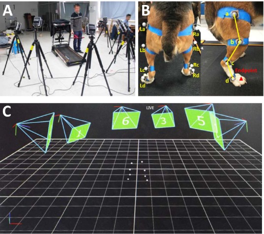
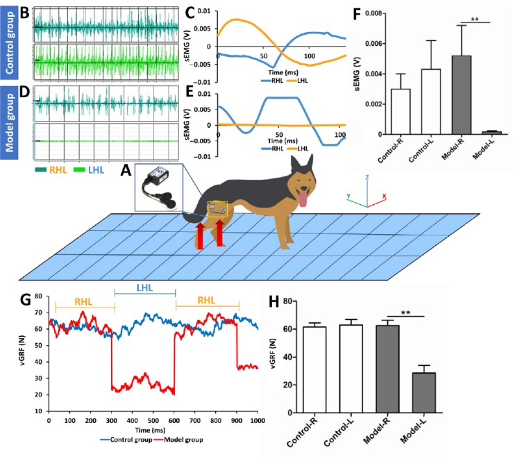
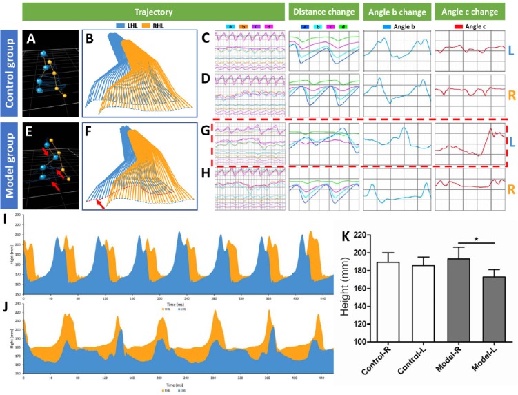
An accurate and effective neurological evaluation is indispensable in the treatment and rehabilitation of traumatic brain injury. However, most of the existing evaluation methods in basic research and clinical practice are not objective or intuitive for assessing the neurological function of big animals, and are also difficult to use to qualify the extent of damage and recovery. In the present study, we established a big animal model of traumatic brain injury by impacting the cortical motor region of beagles. At 2 weeks after successful modeling, we detected neurological deficiencies in the animal model using a series of techniques, including three-dimensional motion capture, electromyogram and ground reaction force. These novel technologies may play an increasingly important role in the field of traumatic brain injury diagnosis and rehabilitation in the future. The experimental protocol was approved by the Animal Care and Use Committee of Logistics University of People's Armed Police Force (approval No. 2017-0006.2).
Keywords: electromyogram; evaluation method; ground reaction force; motion capture; nerve regeneration; neural regeneration; neurological deficiency; traumatic brain injury.
Conflict of interest statement
None
Figures
Similar articles
-
Collagen/heparan sulfate porous scaffolds loaded with neural stem cells improve neurological function in a rat model of traumatic brain injury.Neural Regen Res. 2021 Jun;16(6):1068-1077. doi: 10.4103/1673-5374.300458. Neural Regen Res. 2021. PMID: 33269752 Free PMC article.
-
Collagen scaffold combined with human umbilical cord-mesenchymal stem cells transplantation for acute complete spinal cord injury.Neural Regen Res. 2020 Sep;15(9):1686-1700. doi: 10.4103/1673-5374.276340. Neural Regen Res. 2020. PMID: 32209773 Free PMC article.
-
Delayed inhibition of Nogo-A does not alter injury-induced axonal sprouting but enhances recovery of cognitive function following experimental traumatic brain injury in rats.Neuroscience. 2005;134(3):1047-56. doi: 10.1016/j.neuroscience.2005.04.048. Neuroscience. 2005. PMID: 15979242
-
[Aiming for zero blindness].Nippon Ganka Gakkai Zasshi. 2015 Mar;119(3):168-93; discussion 194. Nippon Ganka Gakkai Zasshi. 2015. PMID: 25854109 Review. Japanese.
-
Considerations for Experimental Animal Models of Concussion, Traumatic Brain Injury, and Chronic Traumatic Encephalopathy-These Matters Matter.Front Neurol. 2017 Jun 1;8:240. doi: 10.3389/fneur.2017.00240. eCollection 2017. Front Neurol. 2017. PMID: 28620350 Free PMC article. Review.
Cited by
-
Pathological significance of tRNA-derived small RNAs in neurological disorders.Neural Regen Res. 2020 Feb;15(2):212-221. doi: 10.4103/1673-5374.265560. Neural Regen Res. 2020. PMID: 31552886 Free PMC article.
-
Three-dimensional-printed collagen/chitosan/secretome derived from HUCMSCs scaffolds for efficient neural network reconstruction in canines with traumatic brain injury.Regen Biomater. 2022 Jun 27;9:rbac043. doi: 10.1093/rb/rbac043. eCollection 2022. Regen Biomater. 2022. PMID: 35855109 Free PMC article.
References
-
- Alizadeh M, Zindl C, Allen MJ, Knapik GG, Fitzpatrick N, Marras WS. MRI cross sectional atlas of normal canine cervical musculoskeletal structure. Res Vet Sci. 2016;109:94–100. - PubMed
-
- Basso DM, Beattie MS, Bresnahan JC. A sensitive and reliable locomotor rating scale for open field testing in rats. J Neurotrauma. 1995;12:1–21. - PubMed
-
- Bederson JB, Pitts LH, Tsuji M, Nishimura MC, Davis RL, Bartkowski H. Rat middle cerebral artery occlusion: evaluation of the model and development of a neurologic examination. Stroke. 1986;17:472–476. - PubMed
-
- Bockstahler B, Kräutler C, Holler P, Kotschwar A, Vobornik A, Peham C. Pelvic limb kinematics and surface electromyography of the vastus lateralis, biceps femoris, and gluteus medius muscle in dogs with hip osteoarthritis. Vet Surg. 2012;41:54–62. - PubMed
LinkOut - more resources
Full Text Sources
