

Ipsilateral motor evoked potentials in a patient with unihemispheric cortical atrophy due to Rasmussen encephalitis
- PMID: 30762014
- PMCID: PMC6404490
- DOI: 10.4103/1673-5374.250581
Ipsilateral motor evoked potentials in a patient with unihemispheric cortical atrophy due to Rasmussen encephalitis
Abstract
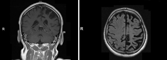
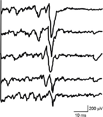
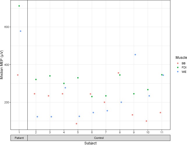
The role of the ipsilaterally descending motor pathways in the recovery mechanisms after unilateral hemispheric damage is still poorly understood. Motor output reorganization was investigated in a 56-year-old male patient with acquired unilateral hemispheric atrophy due to Rasmussen encephalitis. In particular, the ipsilateral corticospinal pathways were explored using focal transcranial magnetic stimulation. In the first dorsal interosseous and wrist extensors muscles, the median amplitudes of the ipsilateral motor evoked potentials induced by transcranial magnetic stimulation in the patient were higher than those of 10 age-matched healthy control subjects. In the biceps brachii muscle, the median amplitudes of the ipsilateral motor evoked potentials were the second largest in the patient compared to the controls. This study demonstrated a reinforcement of ipsilateral motor projections from the unaffected motor cortex to the hemiparetic hand in a subject with acquired unihemispheric cortical damage.
Keywords: Rasmussen encephalitis; cortical atrophy; hemispheric damage; ipsilateral motor evoked potentials; ipsilateral motor pathways; motor cortex; transcranial magnetic stimulation.
Conflict of interest statement
None
Figures
References
-
- Basu AP, Turton A, Lemon RN. Activation of ipsilateral upper limb muscles by transcranial magnetic stimulation. J Physiol. 1994;479:144–145.
-
- Benecke R, Meyer BU, Freund HJ. Reorganisation of descending motor pathways in patients after hemispherectomy and severe hemispheric lesions demonstrated by magnetic brain stimulation. Exp Brain Res. 1991;83:419–426. - PubMed
LinkOut - more resources
Full Text Sources
