Use of a combination strategy to improve neuroprotection and neuroregeneration in a rat model of acute spinal cord injury
- PMID: 30762019
- PMCID: PMC6404491
- DOI: 10.4103/1673-5374.250627
Use of a combination strategy to improve neuroprotection and neuroregeneration in a rat model of acute spinal cord injury
Abstract
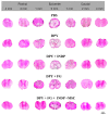
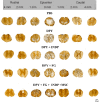
Spinal cord injury is a very common pathological event that has devastating functional consequences in patients. In recent years, several research groups are trying to find an effective therapy that could be applied in clinical practice. In this study, we analyzed the combination of different strategies as a potential therapy for spinal cord injury. Immunization with neural derived peptides (INDP), inhibition of glial scar formation (dipyridyl: DPY), as well as the use of biocompatible matrix (fibrin glue: FG) impregnated with bone marrow mesenchymal stem cells (MSCs) were combined and then its beneficial effects were evaluated in the induction of neuroprotection and neuroregeneration after acute SCI. Sprague-Dawley female rats were subjected to a moderate spinal cord injury and then randomly allocated into five groups: 1) phosphate buffered saline; 2) DPY; 3) INDP + DPY; 4) DPY+ FG; 5) INDP + DPY + FG + MSCs. In all rats, intervention was performed 72 hours after spinal cord injury. Locomotor and sensibility recovery was assessed in all rats. At 60 days after treatment, histological examinations of the spinal cord (hematoxylin-eosin and Bielschowsky staining) were performed. Our results showed that the combination therapy (DPY+ INDP + FG + MSCs) was the best strategy to promote motor and sensibility recovery. In addition, significant increases in tissue preservation and axonal density were observed in the combination therapy group. Findings from this study suggest that the combination theapy (DPY+ INDP + FG + MSCs) exhibits potential effects on the protection and regeneration of neural tissue after acute spinal cord injury. All procedures were approved by the Animal Bioethics and Welfare Committee (approval No. 178544; CSNBTBIBAJ 090812960) on August 15, 2016.
Keywords: fibrin glue; glial scar; mesenchymal stem cells; neural derived peptides; neural regeneration; protective autoimmunity.
Conflict of interest statement
None
Figures


Similar articles
-
Use of a Combination Strategy to Improve Morphological and Functional Recovery in Rats With Chronic Spinal Cord Injury.Front Neurol. 2020 Apr 2;11:189. doi: 10.3389/fneur.2020.00189. eCollection 2020. Front Neurol. 2020. PMID: 32300328 Free PMC article.
-
Immunization with neural derived peptides plus scar removal induces a permissive microenvironment, and improves locomotor recovery after chronic spinal cord injury.BMC Neurosci. 2017 Jan 5;18(1):7. doi: 10.1186/s12868-016-0331-2. BMC Neurosci. 2017. PMID: 28056790 Free PMC article.
-
Motor Recovery after Chronic Spinal Cord Transection in Rats: A Proof-of-Concept Study Evaluating a Combined Strategy.CNS Neurol Disord Drug Targets. 2019;18(1):52-62. doi: 10.2174/1871527317666181105101756. CNS Neurol Disord Drug Targets. 2019. PMID: 30394222
-
Bone marrow mesenchymal stem cells (BMSCs) improved functional recovery of spinal cord injury partly by promoting axonal regeneration.Neurochem Int. 2018 May;115:80-84. doi: 10.1016/j.neuint.2018.02.007. Epub 2018 Feb 16. Neurochem Int. 2018. PMID: 29458076 Review.
-
Schwann cell transplantation for spinal cord injury repair: its significant therapeutic potential and prospectus.Rev Neurosci. 2015;26(2):121-8. doi: 10.1515/revneuro-2014-0068. Rev Neurosci. 2015. PMID: 25581750 Review.
Cited by
-
Pathophysiology of Spinal Cord Injury and Tissue Engineering Approach for Its Neuronal Regeneration: Current Status and Future Prospects.Adv Exp Med Biol. 2023;1409:51-81. doi: 10.1007/5584_2022_731. Adv Exp Med Biol. 2023. PMID: 36038807 Review.
-
Supplementation With Vitamin E, Zinc, Selenium, and Copper Re-Establishes T-Cell Function and Improves Motor Recovery in a Rat Model of Spinal Cord Injury.Cell Transplant. 2022 Jan-Dec;31:9636897221109884. doi: 10.1177/09636897221109884. Cell Transplant. 2022. PMID: 35808825 Free PMC article.
-
Use of Cells, Supplements, and Peptides as Therapeutic Strategies for Modulating Inflammation after Spinal Cord Injury: An Update.Int J Mol Sci. 2023 Sep 11;24(18):13946. doi: 10.3390/ijms241813946. Int J Mol Sci. 2023. PMID: 37762251 Free PMC article. Review.
-
Regional biomechanical characterization of the spinal cord tissue: dynamic mechanical response.Front Bioeng Biotechnol. 2024 Aug 16;12:1439323. doi: 10.3389/fbioe.2024.1439323. eCollection 2024. Front Bioeng Biotechnol. 2024. PMID: 39219623 Free PMC article.
-
Effects of neural stem cell transplantation on the motor function of rats with contusion spinal cord injuries: a meta-analysis.Neural Regen Res. 2020 Apr;15(4):748-758. doi: 10.4103/1673-5374.266915. Neural Regen Res. 2020. PMID: 31638100 Free PMC article.
References
LinkOut - more resources
Full Text Sources

