. 2019 Jun;76(11):2199-2216.
doi: 10.1007/s00018-019-03037-0. Epub 2019 Feb 14.

Nuclear pore protein TPR associates with lamin B1 and affects nuclear lamina organization and nuclear pore distribution

Affiliations

Nuclear pore protein TPR associates with lamin B1 and affects nuclear lamina organization and nuclear pore distribution

Jindřiška Fišerová et al. Cell Mol Life Sci. 2019 Jun.

Abstract

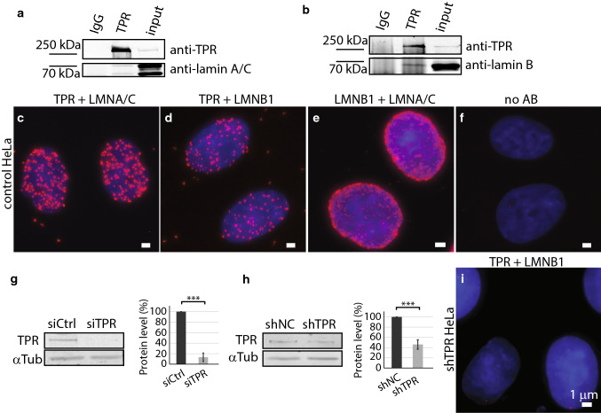
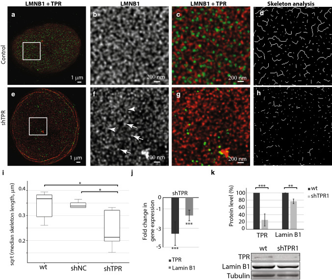
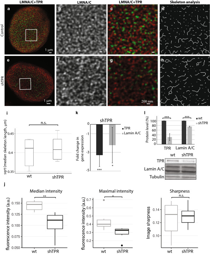
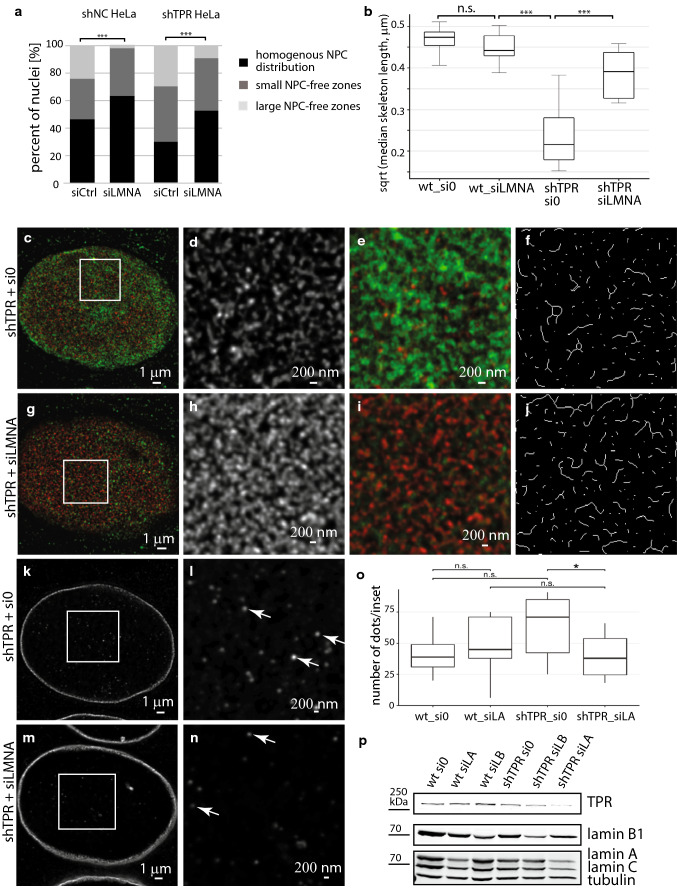
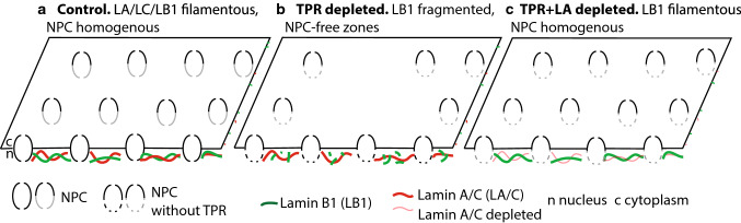
The organization of the nuclear periphery is crucial for many nuclear functions. Nuclear lamins form dense network at the nuclear periphery and play a substantial role in chromatin organization, transcription regulation and in organization of nuclear pore complexes (NPCs). Here, we show that TPR, the protein located preferentially within the nuclear baskets of NPCs, associates with lamin B1. The depletion of TPR affects the organization of lamin B1 but not lamin A/C within the nuclear lamina as shown by stimulated emission depletion microscopy. Finally, reduction of TPR affects the distribution of NPCs within the nuclear envelope and the effect can be reversed by simultaneous knock-down of lamin A/C or the overexpression of lamin B1. Our work suggests a novel role for the TPR at the nuclear periphery: the TPR contributes to the organization of the nuclear lamina and in cooperation with lamins guards the interphase assembly of nuclear pore complexes.

Keywords: Image analysis; Lamina; Lamins; Nuclear pore complex; Nucleus; Super-resolution imaging; TPR; Translocated promoter region.


Conflict of interest statement

The authors declare no competing or financial interests.

Figures

Fig. 1
Fig. 1
TPR co-immunoprecipitates with lamin B1. a Immunoprecipitation of lamin A/C by anti-TPR antibody. b Immunoprecipitation of lamin B1 by anti-TPR antibody. Proximity ligation assay shows the close distance between TPR and lamin A/C (c), TPR and lamin B1 (d), lamin B1 and lamin A/C (e) and negative control with no primary antibody used (f). Western blotting and quantification show the reduction of TPR amount in HeLa cells with siTPR for 48 h: Student t test, T = 12.35, df = 5, P < 0.001 (g) or in stable HeLa cell line expressing shTPR: Student t test, T = 11.01, df = 7, P < 0.001. Graphs represent data from three independent biological replicates. 3.5 × 106 cells were used as a starting material. 10 μg of total protein per each lane was loaded for the analysis (h). i Proximity ligation assay detected reduced abundance of the TPR–lamin B1-positive signal in Hela cells with reduced TPR protein levels by shRNA. Scale bar 1 µm. ***P < 0.001
Fig. 2
Fig. 2
Lamin B1 network is affected by long-term (3 weeks) reduction of TPR amount. The lamin B1 network in control cell nucleus (ad) and in a nucleus with reduced TPR (eh) as visualized by STED. Cells were immunolabeled with rabbit anti-lamin B1 and mouse anti-TPR antibodies. Lamin B1 is shown in red, TPR is depicted in green. a, e Peripheral z-sections show the overview of lamin B1 organization. Insets in a and e correspond to bd and fh, respectively. Lamin B1 forms dense network of randomly organized filaments of similar intensity (b) that appear fragmented and spotted after TPR knock-down (f). Arrows point to brighter lamin B1 spots, arrowheads demarcate lamin B1 filaments of reduced intensity. c, g Merged image of TPR (in green) and lamin B1 (in red). d, h The graphical visualisation of skeleton analysis of b and f, respectively. i The box plots show square root transformed median lengths of skeletonized lamin B1 filaments in wild type (wt) HeLa cells, in cells expressing shNonCoding RNA (shNC) as well as in shTPR cell line. Wt × shTPR: T = 2.44, df = 6.98, P = 0.04; shNC × shTPR: T = 2.79, df = 4.26, P = 0.047; j fold change in mRNA levels of lamin B1 in shTPR cell line. TPR fold change: T = 8.0, df = 5; P < 0.001; LMNB1 fold change: T = 8.0, df = 5, P < 0.001. k Quantification of lamin B1 protein amount after TPR knock-down. TPR protein levels: Student t test: T = 21, df = 8, P < 0.001; LMNB1 protein levels: T = 4.7, df = 8, P < 0.01. Graph represents data from three independent biological replicates analyzed in triplicates. Lower panel shows representative western blots stained with anti-TPR, anti-lamin B1 and anti-tubulin antibodies in control and in shTPR cells. *P < 0.05, **P < 0.01, ***P < 0.001. 15 μg of total protein per each lane was loaded
Fig. 3
Fig. 3
Lamin A/C network is not affected by long-term (3 weeks) reduced TPR amount by stable expression of shTPR. The lamin A/C network in control HeLa cell nucleus (ad) and in a cell nucleus with reduced TPR amount (eh) as visualized by STED. Cells were immunolabeled with mouse anti-lamin A/C (in red) and rabbit anti-TPR (in green). a, e Peripheral z-sections show the overview of lamin A/C organization. Insets in a and e correspond to bd and fh, respectively. Lamin A/C forms dense network of randomly organized filaments (b) that does not change in response to reduced TPR amount (f). c, g Merged image of lamin A/C (in red) and TPR (in green). d, h The graphical visualisation of skeleton analysis of the insets b and f, respectively. i Box plot shows square root transformed median lengths of skeletonized lamin A/C filaments in wild-type (wt) HeLa cells and in shTPR. Wt × shTPR: Student t test: T = − 0.61, df = 9.91, P = 0.6. j Comparison of descriptive statistics of median and maximal intensity and sharpness of the images. Intensity in sharpness was scaled to fit the range of 0–1. TPR depletion decreased both median (Wilcoxon exact test, W = 35.5, P = 0.004) and maximal (W = 33, P = 0.015) fluorescent intensity. k Fold change in mRNA levels of lamin A/C in shTPR cell line. TPR fold change: T = 7.2, df = 6, P < 0.001; LMNA/C fold change: T = 3.1, df = 6, P < 0.05. l Quantification of lamin A/C protein amount after TPR knock-down. TPR protein levels: T = 20.6, df = 8, P < 0.001; LMNA/C protein levels: T = 8.3, df = 8, P < 0.001. Graph represents data from three independent biological replicates analyzed in triplicates. Lower panel shows representative western blots stained with anti-TPR, anti-lamin A/C and anti-tubulin antibodies in control and in shTPR cells. ***P < 0.001, **P < 0.01, *P < 0.05, n.s. non-significant. 15 μg of total protein per each lane was loaded
Fig. 4
Fig. 4
A distribution of NPCs is affected in TPR-depleted HeLa cells (shTPR HeLa cell line). ac The distribution of NPCs in control HeLa cells as viewed by immunostaining with anti-Nup62 antibody and wide-field Leica DM6000 microscope. a Homogenous distribution. b Small NPC-free zones. c Large NPC-free zones comprehend more than 1/10 of nuclear surface. d The ratio of homogenous distribution, small and large NPC-free zones in control wild-type S-G2 cells (wt), in cells stably expressing shRNA targeting non-coding regions (shNC) and TPR (shTPR cells). In TPR-depleted cells, the number of nuclei with NPC-free zones increased significantly (χ2 = 11.41, df = 2, P < 0.001, Chi squared test), **P < 0.01, ***P < 0.001, n.s. not significant. e The ratio of homogenous, small and large NPC-free zones in control wild type cells (wt) and in cells stably expressing shRNA targeting non-coding regions (shNC) and TPR (shTPR) synchronized in G1/S and in G2 phase of the cell cycle. The number of NPC-free zones significantly increased in TPR-depleted cells (***P < 0.001, n.s. not significant). fi Recruitment of nucleoporins Nup107, Nup133 and central FG-Nups into NPC-free zones is impaired in TPR-depleted cells. f The ratio of distribution of NPC-free zones in shNC and shTPR cells stained with Nup153 antibody (χ2 = 7.08, df = 2, P = 0.029). g The ratio of homogenous distribution, small and large NPC-free zones in shNC and shTPR cells transfected with GFP–Nup107 construct (χ2 = 9.713, df = 2, P = 0.008). h The ratio of homogenous distribution, small and large NPC-free zones in shNC and shTPR cells overexpressing GFP–Nup133 (χ2 = 23.855, df = 2, P < 0.001). i The ratio of distribution of NPC-free zones in shNC and shTPR cells stained with antibody recognizing central FG-Nups (χ2 = 138.5, df = 2, P < 0.0001)
Fig. 5
Fig. 5
Lamin B1 levels influence NPC distribution in HeLa cell population with reduced TPR amount. a Knock-down of lamin B1 prevents homogenous distribution of NPCs in HeLa cells expressing RNA against non-coding genomic region (shNC) (test of interaction in Poisson mixed-effects model, χ2 = 9.65, df = 2, P < 0.05). Reduction of lamin B1 in shTPR cells slightly increases the numbers of nuclei with NPC-free zones (χ2 = 4.79, df = 2, P < 0.1). b GFP–lamin B1 overexpression did not affect the NPC distribution of the entire population of control HeLa cells. Overexpression of GFP–lamin B1 partially rescued the normal proportions of homogenous NPC distributions in shTPR cells (χ2 = 13.39, df = 2, P < 0.01). ***P < 0.001, *P < 0.05, P < 0.1, n.s. not significant
Fig. 6
Fig. 6
Reduction of lamin A levels can rescue the NPC distribution as well as an organization of lamin B1 peripheral network in shTPR HeLa cells. a Knock-down of lamin A favors homogenous NPC distribution in shTPR cells (test of interaction in Poisson mixed-effects model; shNC: χ2 = 0.39, df = 2, P = 0.82; shTPR: χ2 = 8.57, df = 2, P < 0.05). bj Reduction of lamin A levels in shTPR HeLa cells rescues the lamin B1 network organization. b Box plot shows square root transformed median lengths of the skeletonized lamin B1 filaments in control cells as well as in cells with reduced TPR and lamin A levels. The lamin A knock-down results in elongation of lamin B1 filaments in shTPR cells in comparison to shTPR only. Wt_si0 × wt_siLMNA: Student t test: T = 1.1, df = 14, P = 0.29; wt_siLMNA × shTPR_si0: T = 8.6, df = 14, P < 0.001; shTPR_si0 × shTPR_siLMNA: T = − 5.1, df = 16, P < 0.001. The lamin B1 network in cell with reduced TPR levels (cf) and in cell with reduced both, TPR and lamin A amount (gj) as visualized by STED. The cells were immunostained with mouse anti-lamin A/C (shown in green) and rabbit anti-lamin B1 (shown in red). c, g Peripheral z-sections show the overview of lamin A/C//B1 organization. Insets in c and g correspond to df and hj, respectively. Lamin B1 appears fragmented in shTPR cells (d) whereas it seems more polymerized in shTPR cells with reduced lamin A levels (h). e, i Merged images of lamin A/C and lamin B1. f, j The graphical visualisation of skeleton analysis of the insets d and h, respectively. kn STED images of immunofluorescently detected lamin B1 in central z-sections of shTPR cells (k, l) and shTPR cells with reduced lamin A levels (m, n). Insets in k and m correspond to l and n, respectively. Arrows in l and n point to the intranuclear lamin B1 dots. o Analysis of number of lamin B1 dots in central z-sections of STED images of HeLa nuclei immunolabeled with lamin A/C and lamin B1. The number of lamin B1 intranuclear dots decreased significantly under co-depletion of TPR and lamin A (Wilcoxon exact test, W = 48.5, P = 0.045). p Western blotting of lamin levels in control cells and in cells with reduced TPR (shTPR), lamin A (siLA) and lamin B1 (siLB) levels shows that lamin B1 amount did not increase in response to lamin A reduction in shTPR cells. *P < 0.05, ***P < 0.001, n.s. non-significant. 3.5 × 106 cells were used as a starting material. 20 μg of total protein per each lane was loaded
Fig. 7
Fig. 7
A scheme showing interdependence of lamin A/C, lamin B1 and TPR organization and NPC distribution in HeLa cells. a In control cells, lamin A/C and lamin B1 are fully polymerized, TPR forms nuclear baskets of NPCs that are homogenously distributed at the nuclear envelope. b Long-term TPR depletion results in lamin B1 (but not lamin A/C) network fragmentation, reduction of lamin B1 levels and the presence of NPC-free zones. c Co-depletion of TPR and lamin A restores both lamin B1 network organization and NPC homogenous distribution

Similar articles

Cited by

References

    1. Gruenbaum Y, Foisner R. Lamins: nuclear intermediate filament proteins with fundamental functions in nuclear mechanics and genome regulation. Annu Rev Biochem. 2015;84:131–164. doi: 10.1146/annurev-biochem-060614-034115. - DOI - PubMed
    1. Naetar N, Ferraioli S, Foisner R. Lamins in the nuclear interior—life outside the lamina. J Cell Sci. 2017;130(13):2087–2096. doi: 10.1242/jcs.203430. - DOI - PubMed
    1. de Leeuw R, Gruenbaum Y, Medalia O. Nuclear lamins: thin filaments with major functions. Trends Cell Biol. 2017 doi: 10.1016/j.tcb.2017.08.004. - DOI - PubMed
    1. Schermelleh L, Carlton PM, Haase S, Shao L, Winoto L, Kner P, Burke B, Cardoso MC, Agard DA, Gustafsson MG, Leonhardt H, Sedat JW. Subdiffraction multicolor imaging of the nuclear periphery with 3D structured illumination microscopy. Science. 2008;320(5881):1332–1336. doi: 10.1126/science.1156947. - DOI - PMC - PubMed
    1. Shimi T, Kittisopikul M, Tran J, Goldman AE, Adam SA, Zheng Y, Jaqaman K, Goldman RD. Structural organization of nuclear lamins A, C, B1, and B2 revealed by superresolution microscopy. Mol Biol Cell. 2015;26(22):4075–4086. doi: 10.1091/mbc.E15-07-0461. - DOI - PMC - PubMed

MeSH terms