

Alpha protons as NMR probes in deuterated proteins
- PMID: 30762170
- PMCID: PMC6441447
- DOI: 10.1007/s10858-019-00230-y
Alpha protons as NMR probes in deuterated proteins
Abstract
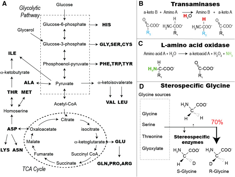
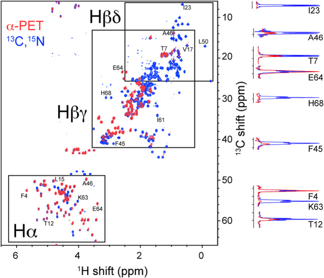
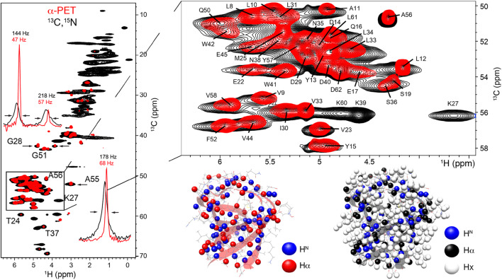
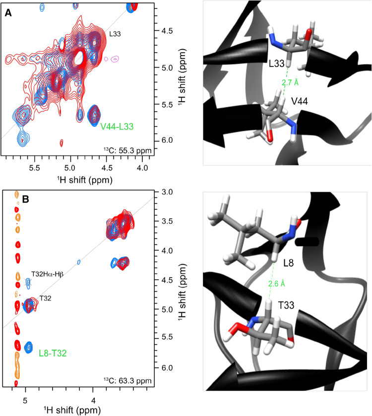
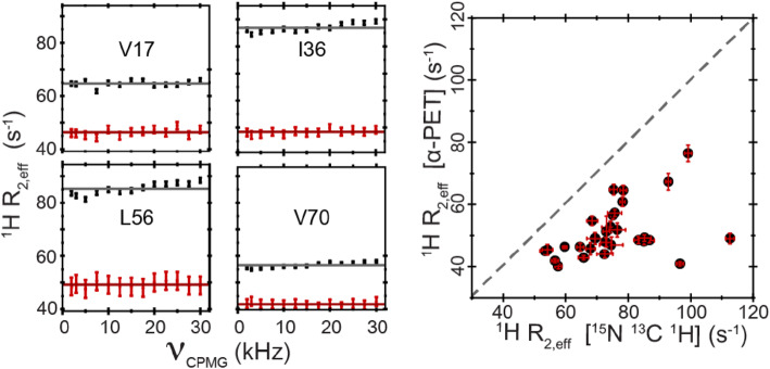
We describe a new labeling method that allows for full protonation at the backbone Hα position, maintaining protein side chains with a high level of deuteration. We refer to the method as alpha proton exchange by transamination (α-PET) since it relies on transaminase activity demonstrated here using Escherichia coli expression. We show that α-PET labeling is particularly useful in improving structural characterization of solid proteins by introduction of an additional proton reporter, while eliminating many strong dipolar couplings. The approach benefits from the high sensitivity associated with 1.3 mm samples, more abundant information including Hα resonances, and the narrow proton linewidths encountered for highly deuterated proteins. The labeling strategy solves amide proton exchange problems commonly encountered for membrane proteins when using perdeuteration and backexchange protocols, allowing access to alpha and all amide protons including those in exchange-protected regions. The incorporation of Hα protons provides new insights, as the close Hα-Hα and Hα-HN contacts present in β-sheets become accessible, improving the chance to determine the protein structure as compared with HN-HN contacts alone. Protonation of the Hα position higher than 90% is achieved for Ile, Leu, Phe, Tyr, Met, Val, Ala, Gln, Asn, Thr, Ser, Glu, Asp even though LAAO is only active at this degree for Ile, Leu, Phe, Tyr, Trp, Met. Additionally, the glycine methylene carbon is labeled preferentially with a single deuteron, allowing stereospecific assignment of glycine alpha protons. In solution, we show that the high deuteration level dramatically reduces R2 relaxation rates, which is beneficial for the study of large proteins and protein dynamics. We demonstrate the method using two model systems, as well as a 32 kDa membrane protein, hVDAC1, showing the applicability of the method to study membrane proteins.
Keywords: Isotopic labeling; L-Amino acid oxidase; Membrane proteins; NMR; Structural restraints; Transamination.
Figures


References
-
- Agarwal V, Reif B. Residual methyl protonation in perdeuterated proteins for multi-dimensional correlation experiments in MAS solid-state NMR spectroscopy. J Magn Reson. 2008;194:16–24. - PubMed
-
- Agarwal V, Linser R, Fink U, Faelber K, Reif B. Identification of hydroxyl protons, determination of their exchange dynamics, and characterization of hydrogen bonding in a microcrystallin protein. J Am Chem Soc. 2010;132:3187–3195. - PubMed
-
- Akbey Ü, Lange S, Franks WT, Linser R, Rehbein K, Diehl A, Van Rossum BJ, Reif B, Oschkinat H. Optimum levels of exchangeable protons in perdeuterated proteins for proton detection in MAS solid-state NMR spectroscopy. J Biomol NMR. 2010;46:67–73. - PubMed
-
- Andreas LB, Le T, Jaudzems K, Pintacuda G. High-resolution proton-detected NMR of proteins at very fast MAS. J Magn Reson. 2015;253:36–49. - PubMed
-
- Andreas LB, Jaudzems K, Stanek J, Lalli D, Bertarello A, Le Marchand T, Cala-De Paepe D, Kotelovica S, Akopjana I, Knott B, Wegner S, Engelke F, Lesage A, Emsley L, Tars K, Herrmann T, Pintacuda G. Structure of fully protonated proteins by proton-detected magic-angle spinning NMR. Proc. Natl. Acad. Sci. 2016;113:9187–9192. - PMC - PubMed
MeSH terms
Substances
Grants and funding
LinkOut - more resources
Full Text Sources
Miscellaneous
