

Case Reports
doi: 10.1213/XAA.0000000000000977.
Localized Corticosteroid Injections for Malignant Joint Pain in the Oncologic Population: A Case Series
Affiliations
- PMID: 30762588
- PMCID: PMC9476896
- DOI: 10.1213/XAA.0000000000000977
Item in Clipboard
Case Reports
Localized Corticosteroid Injections for Malignant Joint Pain in the Oncologic Population: A Case Series
A A Pract.
.
Abstract
Pain is a common issue that is present in cancer survivors as well as those with active malignant processes. Despite opioid analgesics and adjuvant therapies such as systemic corticosteroids, many patients have persistent localized pain. We describe a case series of 3 cancer patients who have concurrent hip- and greater trochanteric-related pain. We performed a single-insertion-site, ultrasound-guided injection to target both the intra-articular hip and greater trochanteric bursa for each patient. All patients reported an improvement in pain symptoms and function with no major complications. Targeted corticosteroid injections provide a potential for relief of malignant joint pain.
Figures

(a) Example of coronal oblique probe and needle positioning of the acetabular and femoral head/neck junction. (b) Needle direction for intra-articular hip joint injection. (c) Needle direction for greater trochanteric bursa injection.

(a) Example of coronal oblique probe and needle positioning of the acetabular and femoral head/neck junction. (b) Needle direction for intra-articular hip joint injection. (c) Needle direction for greater trochanteric bursa injection.

(a) Example of coronal oblique probe and needle positioning of the acetabular and femoral head/neck junction. (b) Needle direction for intra-articular hip joint injection. (c) Needle direction for greater trochanteric bursa injection.
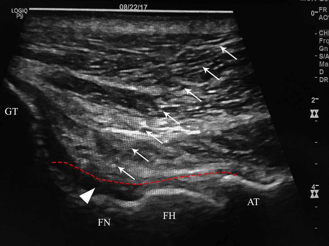
(a, b) Ultrasound coronal oblique view of the acetabular and femoral head/neck junction with and without labels. (c) Example of a long-axis approach of the intra-articular hip joint. (d) Example of a long-axis approach of the greater trochanteric bursa. White arrows indicate needle. White arrowhead indicates needle tip. Green indicates gluteus medius. Orange indicates gluteus minimus. Purple indicates iliofemoral ligament. GT greater trochanter, FN femoral neck, FH femoral head, AT acetabulum. Dotted red line indicates the hip joint capsule. Dotted blue line indicates greater trochanteric bursa.

(a, b) Ultrasound coronal oblique view of the acetabular and femoral head/neck junction with and without labels. (c) Example of a long-axis approach of the intra-articular hip joint. (d) Example of a long-axis approach of the greater trochanteric bursa. White arrows indicate needle. White arrowhead indicates needle tip. Green indicates gluteus medius. Orange indicates gluteus minimus. Purple indicates iliofemoral ligament. GT greater trochanter, FN femoral neck, FH femoral head, AT acetabulum. Dotted red line indicates the hip joint capsule. Dotted blue line indicates greater trochanteric bursa.

(a, b) Ultrasound coronal oblique view of the acetabular and femoral head/neck junction with and without labels. (c) Example of a long-axis approach of the intra-articular hip joint. (d) Example of a long-axis approach of the greater trochanteric bursa. White arrows indicate needle. White arrowhead indicates needle tip. Green indicates gluteus medius. Orange indicates gluteus minimus. Purple indicates iliofemoral ligament. GT greater trochanter, FN femoral neck, FH femoral head, AT acetabulum. Dotted red line indicates the hip joint capsule. Dotted blue line indicates greater trochanteric bursa.

(a, b) Ultrasound coronal oblique view of the acetabular and femoral head/neck junction with and without labels. (c) Example of a long-axis approach of the intra-articular hip joint. (d) Example of a long-axis approach of the greater trochanteric bursa. White arrows indicate needle. White arrowhead indicates needle tip. Green indicates gluteus medius. Orange indicates gluteus minimus. Purple indicates iliofemoral ligament. GT greater trochanter, FN femoral neck, FH femoral head, AT acetabulum. Dotted red line indicates the hip joint capsule. Dotted blue line indicates greater trochanteric bursa.

(a, b) Case 1, CT pelvis reveals posterior osteophytes on the left femoral head (open arrowhead, a) and left retroperitoneal soft tissue iliac mass (open arrowhead, b) (c) Case 2, MRI hip shows a right hip intra-articular lesion (open arrowhead) (d) Case 3, CT pelvis demonstrates a right greater trochanteric lytic lesion (open arrowhead) with posterior sclerosis (asterisk).

(a, b) Case 1, CT pelvis reveals posterior osteophytes on the left femoral head (open arrowhead, a) and left retroperitoneal soft tissue iliac mass (open arrowhead, b) (c) Case 2, MRI hip shows a right hip intra-articular lesion (open arrowhead) (d) Case 3, CT pelvis demonstrates a right greater trochanteric lytic lesion (open arrowhead) with posterior sclerosis (asterisk).

(a, b) Case 1, CT pelvis reveals posterior osteophytes on the left femoral head (open arrowhead, a) and left retroperitoneal soft tissue iliac mass (open arrowhead, b) (c) Case 2, MRI hip shows a right hip intra-articular lesion (open arrowhead) (d) Case 3, CT pelvis demonstrates a right greater trochanteric lytic lesion (open arrowhead) with posterior sclerosis (asterisk).

(a, b) Case 1, CT pelvis reveals posterior osteophytes on the left femoral head (open arrowhead, a) and left retroperitoneal soft tissue iliac mass (open arrowhead, b) (c) Case 2, MRI hip shows a right hip intra-articular lesion (open arrowhead) (d) Case 3, CT pelvis demonstrates a right greater trochanteric lytic lesion (open arrowhead) with posterior sclerosis (asterisk).
References
-
- White P, Arnold R, Bull J, Cicero B. The Use of Corticosteroids as Adjuvant Therapy for Painful Bone Metastases: A Large Cross-Sectional Survey of Palliative Care Providers. American Journal of Hospice and Palliative Medicine. 2016;35(1):151–158. - PubMed
-
- Roodman GD. Mechanisms of Bone Metastasis. New England Journal of Medicine. 2004;350(16):1655–1664. - PubMed
-
- Twycross R, Harcourt J, Bergl S. A survey of pain in patients with advanced cancer. Journal of Pain and Symptom Management. 1996;12(5):273–282. - PubMed
-
- Tannock I, Gospodarowicz M, Meakin W, Panzarella T, Stewart L, Rider W. Treatment of metastatic prostatic cancer with low-dose prednisone: evaluation of pain and quality of life as pragmatic indices of response. Journal of Clinical Oncology. 1989;7(5):590–597. - PubMed
Publication types
MeSH terms
Substances
Grants and funding
LinkOut - more resources
Full Text Sources
Medical
