Review
. 2019 Mar;39(1):94-102.
doi: 10.1097/WNO.0000000000000766.

Neurobiology of Photophobia

Affiliations
Review

Neurobiology of Photophobia

Rami Burstein et al. J Neuroophthalmol. 2019 Mar.

Abstract

Background: Photophobia is commonly associated with migraine, meningitis, concussion, and a variety of ocular diseases. Advances in our ability to trace multiple brain pathways through which light information is processed have paved the way to a better understanding of the neurobiology of photophobia and the complexity of the symptoms triggered by light.

Purpose: The purpose of this review is to summarize recent anatomical and physiological studies on the neurobiology of photophobia with emphasis on migraine.

Recent findings: Observations made in blind and seeing migraine patients, and in a variety of animal models, have led to the discovery of a novel retino-thalamo-cortical pathway that carries photic signal from melanopsinergic and nonmelanopsinergic retinal ganglion cells (RGCs) to thalamic neurons. Activity of these neurons is driven by migraine and their axonal projections convey signals about headache and light to multiple cortical areas involved in the generation of common migraine symptoms. Novel projections of RGCs into previously unidentified hypothalamic neurons that regulate parasympathetic and sympathetic functions have also been discovered. Finally, recent work has led to a novel understanding of color preference in migraine-type photophobia and of the roles played by the retina, thalamus, and cortex.

Summary: The findings provide a neural substrate for understanding the complexity of aversion to light in patients with migraine and neuro-ophthalmologic other disorders.


Conflict of interest statement

Conflict of Interest: Dr. Burstein received grants support for hisa studies on migraine pathophysiology from Teva, Allergan, Dr. Reddy, and Trigemina; he also serves as a consultant to Alder Biopharm, Allergan, Amgen, Autonomic Technologies Inc., Avanir, Biohaven, Depomed, Dr. Reddy, Electrocore, Johnson and Johnson, Neurolief, Percept, Pernix, Strategic Science and Technologies, Teva, Theranica, Trigemina. Beth Israel Deaconess Medical center and Dr. Burstein hold a provisional patent on the use of narrow band green light for the treatment of photophobia in migraine.

Figures

Figure 1.
Figure 1.
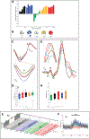
Neuronal pathways that mediate exacerbation of headache by light. Proposed mechanism for exacerbation of migraine headache by light through the convergence of the photic signals from the retina and nociceptive signals from the meninges on the same thalamic neurons that project to the somatosensory cortices. Red depicts the trigeminovascular pathway. Blue depicts visual pathway from the retina to the posterior thalamus. Inserts show distribution of trigeminovascular axons in the somatosensory cortex (top-right), monosynaptic connections between axons of RGC and dendrites of thalamic trigeminovascular neurons (bottom-left) and light effect on activation pattern of a thalamic trigeminovascular neuron (bottom-left). Abbreviations: RGC, retinal ganglion cells; ipRGC, intrinsically-photosensitive retinal ganglion cells; TG, trigeminal ganglion; Sp5, spinal trigeminal nucleus; LP, lateral posterior nucleus; Pul, pulvinar; S1, primary somatosensory cortex; S2 secondary somatosensory cortex. Adapted from [7,14,50,51]
Figure 2.
Figure 2.
Neuronal pathways that mediate common migraine associated symptoms. Proposed mechanism for initiation or exacerbation of common migraine-associated symptoms by light during migraine through convergence of nociceptive signals from the meninges on thalamic neurons that project to the motor (M), parietal association (PtA), auditory (Au), retro-splenius (RS), olfactory (ECT) and visual (V) cortices. Inserts (top-right) show distribution of trigeminovascular axons in the parietal association, retro-splenius and visual cortices. Red depicts the trigeminovascular pathway. Blue depicts visual pathway from the retina to the visual cortex. Additional Abbreviations: as in Fig. 1). Adapted from [7,14,50,51]
Figure 3.
Figure 3.
Proposed mechanism for color preference in migraine-type photophobia. A-B. Effects of color on pain rating and headache location. C. Chromatic electroretinographies (ERGs) recorded in 43 migraine patients. Shown in the figure are superimposed means of standard light-adapted 30 Hz flickering ERG waveforms averaged (± SEM) across patients in response to each color of light. These light-adapted flickering ERG waveforms illustrate that blue, red and white lights generated significantly larger a-wave amplitudes as compared to green light (enlarged view in inset) (p<0.0001). D. Boxplot illustrating median, 95% CI, interquartile range, and observations below and above the 25th and 75th percentile, respectively. Asterisks depict the significantly greater a-wave amplitude induced by blue, red and white lights compared to green. E. 3D bar graphs illustrating firing frequency (i.e., raw data expressed in mean spikes/sec, bin size=1 sec) of 37 posterior hypothalamus neurons to 44, 59, 58 and 59 cycles (dark-light-dark, 1-min each) of photic retinal stimulation with white, blue, green and red lights, respectively. Neurons whose activity in the light increased by >2SD over baseline (i.e., in the dark), are marked as responders (resp) and shown on the right side of each collections of color-selective bar graphs. The data were collected using sophisticated 3-tetrode in vivo multi-unit recording technique. F. Differential responses of trigeminovascular and non-trigeminovascular thalamic neurons to photic stimulation with white, blue, green and red lights. G. Chromatic visual evoked potentials (VEPs) recorded in 28 migraine patients. Shown in the figure are superimposed means of standard flash VEP waveforms averaged (± SEM) across patients in response to 1,792 flashes (64 flashes per patient, 1 s inter-stimulus interval) of each color of light. The superimposed means of these flash VEPs demonstrate that blue, red and amber generated significantly larger P2 amplitude (enlarged view in inset) as compared to green (p<0.02). In contrast, no differences were found in the amplitudes of the N2 waves. H. Boxplot illustrating median (thick horizontal white line), 95% CI (thin dotted horizontal lines), interquartile range (25th-75th percentile; lower and upper box boundaries) and observations below and above the 25th and 75th percentile, respectively (individual dots). Asterisk depicts the significantly smaller P2-wave amplitude induced by the green light compared to red, blue and amber. Adapted from [28].
Figure 4.
Figure 4.
Neuronal pathways that mediate color preference in migraine-type photophobia. Such pathways involve sequential activation of cone photoreceptors, post-cone retinal pathways, cone-opponent retinal ganglion cells, relauy posterior thalamic neurons (located in the pulvinar, lateral posterior and posterior nuclei), and double-opponent neurons in V1, V2 and V4. Abbreviations, as in Fig. 1. Adapted from [50,51]
Figure 5.
Figure 5.
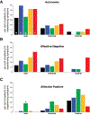
Aversion to light. A. Proportion of migraine patients and control subjects experiencing autonomic responses to white, blue, green, amber, and red lights. B. Proportion of migraine patients and control subjects experiencing negative emotions in response to white, blue, green, amber, and red lights. C. Proportion of migraine patients and control subjects experiencing positive emotions in response to white, blue, green, amber, and red lights. Adapted from [10].
Figure 6.
Figure 6.
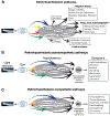
Neuronal pathways that mediate general aversion to light. Proposed pathways for modulation of autonomic responses, hypothalamic functions, and emotions by light. A. Pathways for inductions of symptoms mediated by dopamine, orexin, histamine, melanin concentrating hormone, oxytocin and vasopressin. B. Pathways for induction of symptoms associated with activation of the parasympathetic system. C. Pathways for induction of symptoms associated with activation of the sympathetic system. Adapted from [10].

Similar articles

Cited by

References

    1. Lebensohn JE: Photophobia: mechanism and implications. Am J Ophthalmol 1951, 34:1294–1300. - PubMed
    1. Kawasaki A, Purvin VA: Photophobia as the presenting visual symptom of chiasmal compression. J Neuroophthalmol 2002, 22:3–8. - PubMed
    1. Aurora SK, Cao Y, Bowyer SM, Welch KM: The occipital cortex is hyperexcitable in migraine: experimental evidence. Headache 1999, 39:469–476. - PubMed
    1. Lamonte M, Silberstein SD, Marcelis JF: Headache associated with aseptic meningitis. Headache 1995, 35:520–526. - PubMed
    1. Welty TE, Horner TG: Pathophysiology and treatment of subarachnoid hemorrhage. Clin Pharm 1990, 9:35–39. - PubMed

Publication types

MeSH terms