

Head trauma complicated with primary cranial vault lymphoma: A case report
- PMID: 30762761
- PMCID: PMC6407994
- DOI: 10.1097/MD.0000000000014465
Head trauma complicated with primary cranial vault lymphoma: A case report
Abstract
Rationale: Primary cranial vault lymphoma (PCVL) is an extremely rare extranodal lymphoma in the skull. This case study investigates the clinical features, so as to improve the understanding of the diagnosis and therapy.
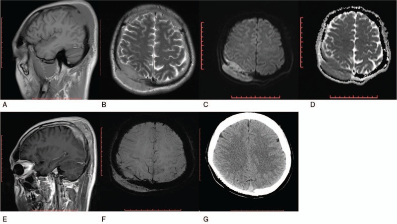
Patient concerns: A 31-year-old male presented painful scalp mass at the site of 1-month-old head trauma.
Diagnosis: The final diagnosis was plasma cell lymphoma, which is a rare subtype of diffuse large B-cell lymphoma based on biopsy and immunohistochemistry findings.
Interventions: The patient received total tumor resection in combination with chemotherapy OUTCOMES:: The patient survived without signs of systemic dissemination for 12 months after surgery at the time of last follow-up.
Lessons: Trauma may be one of the factors that induce PCVL. The final diagnosis of PCVL depends on pathology and immunohistochemistry findings. A combined treatment of surgery, chemotherapy, and radiotherapy can achieve favorable outcomes.
Conflict of interest statement
The authors have no conflicts of interest to disclose.
Figures

Similar articles
-
Primary cranial vault lymphoma with brain infiltration: case report and review of the literature.Br J Neurosurg. 2012 Oct;26(5):756-8. doi: 10.3109/02688697.2012.665515. Epub 2012 Mar 31. Br J Neurosurg. 2012. PMID: 22463812 Review.
-
Diffuse large B cell lymphoma of the cranial vault: two case reports.Brain Tumor Pathol. 2015 Oct;32(4):275-80. doi: 10.1007/s10014-015-0225-5. Epub 2015 Jul 16. Brain Tumor Pathol. 2015. PMID: 26177806
-
Primary diffuse large B-cell lymphoma of the dura mater and cranial vault. Case report and literature review.Neurosurg Focus. 2006 Nov 15;21(5):E10. doi: 10.3171/foc.2006.21.5.11. Neurosurg Focus. 2006. PMID: 17134112 Review.
-
Multiple myeloma complicated by skull plasmacytoma discovered after head injury.J Integr Neurosci. 2021 Jun 30;20(2):459-462. doi: 10.31083/j.jin2002048. J Integr Neurosci. 2021. PMID: 34258947
-
Contiguous scalp, skull, and epidural Hodgkin's disease.Surg Neurol. 1984 Feb;21(2):182-4. doi: 10.1016/0090-3019(84)90339-2. Surg Neurol. 1984. PMID: 6701756
Cited by
-
Characteristics of cranial vault lymphoma from a systematic review of the literature.Surg Neurol Int. 2022 Jun 3;13:231. doi: 10.25259/SNI_28_2022. eCollection 2022. Surg Neurol Int. 2022. PMID: 35855149 Free PMC article. Review.
-
Primary Cranial Vault Lymphoma Extending between Subcutaneous Tissue and Brain Parenchyma without Skull Destruction after Mild Head Trauma: A Case Report and Literature Review.Case Rep Oncol. 2021 Jul 15;14(2):1118-1123. doi: 10.1159/000516272. eCollection 2021 May-Aug. Case Rep Oncol. 2021. PMID: 34413742 Free PMC article.
References
-
- Scuotto A, Rotondo M, Conforti R. Primary lymphoma of the skull base. Eur J Radiol Extra 2008;66:81–3.
-
- Wang L, Lin S, Zhang J, et al. Primary non-Hodgkin's lymphoma of the skull base: a case report and literature review. Clin Neurol Neurosurg 2013;115:237–40. - PubMed
-
- El Asri AC, Akhaddar A, Baallal H, et al. Primary lymphoma of the cranial vault: case report and a systematic review of the literature. Acta Neurochir (Wien) 2012;154:257–65. - PubMed
-
- Tomaszek DE, Tyson GW, Stang P, et al. Contiguous scalp, skull, and epidural Hodgkin's disease. Surg Neurol 1984;21:182–4. - PubMed
Publication types
MeSH terms
LinkOut - more resources
Full Text Sources
Medical
