Intravascular large B-cell lymphoma presenting with hearing loss and dizziness: A case report
- PMID: 30762766
- PMCID: PMC6407998
- DOI: 10.1097/MD.0000000000014470
Intravascular large B-cell lymphoma presenting with hearing loss and dizziness: A case report
Abstract
Rationale: Intravascular large B-cell lymphoma (IVLBCL) is a type of malignant lymphoma in which neoplastic B cells proliferate selectively within the lumina of small- and medium-sized vessels. Patients with IVLBCL frequently develop neurological manifestations during their disease course. Patients are known to often develop various neurological manifestations, but there are only a few reports of IVLBCL whose initial symptoms are deafness and/or disequilibrium.
Patient concerns: A 66-year-old Japanese man was provisionally diagnosed with sudden sensorineural hearing loss. Administration of prednisolone did not improve his symptoms, and then he experienced amaurosis fugax. Magnetic resonance imaging (MRI) showed multiple brain infarcts, so he was administered antithrombotic drugs. Nevertheless, he experienced recurrent strokes, became irritable, and had visual hallucinations. He was emergently admitted to our hospital with disturbance of consciousness.
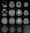
Diagnosis: Blood tests showed elevation of lactose dehydrogenase and soluble interleukin-2 receptor. Cranial MR diffusion-weighted imaging showed multiple lesions bilaterally in the cerebral white matter and cortex, posterior limbs of the internal capsule, and cerebellar hemispheres, which were hypointense on apparent diffusion coefficient maps. Hyperintense lesions were detected bilaterally in the cerebral white matter and basal ganglia on both T2-weighted imaging and fluid-attenuated inversion recovery imaging. Contrast-enhanced brain MRI demonstrated contrast-enhancing high-signal lesions along the cerebral cortex. Brain biopsy revealed a diagnosis of IVLBCL.
Interventions: The patient could not receive chemotherapy because of his poor general condition. Therefore, we administered high-dose methylprednisolone (mPSL) pulse therapy.
Outcomes: There was little improvement in consciousness levels after the high-dose mPSL pulse therapy. On the forty-ninth day of hospitalization, he was transferred to another hospital to receive supportive care.
Lessons: IVLBCL should be regarded as an important differential diagnosis of hearing loss and dizziness. Most importantly, if the symptoms are fluctuant and steroid therapy is not effective, biopsy should be considered as early as possible.
Conflict of interest statement
The authors report no conflicts of interest.
Figures


Similar articles
-
[A case of intravascular large B-cell lymphoma manifested as lacunar infarction].Rinsho Shinkeigaku. 2022 Jun 24;62(6):492-495. doi: 10.5692/clinicalneurol.cn-001704. Epub 2022 May 28. Rinsho Shinkeigaku. 2022. PMID: 35644581 Japanese.
-
Case report: Intravascular large B cell lymphoma mimicking acute demyelinating encephalomyelitis after SARS-CoV-2 reinfection: diagnostic value of advanced MRI techniques and the literature review with the assistance of ChatGPT.Front Immunol. 2024 Nov 14;15:1478163. doi: 10.3389/fimmu.2024.1478163. eCollection 2024. Front Immunol. 2024. PMID: 39611151 Free PMC article. Review.
-
[Intravascular large B-cell lymphoma with multiple intracranial hemorrhages diagnosed by brain biopsy].Rinsho Shinkeigaku. 2020 Mar 31;60(3):206-212. doi: 10.5692/clinicalneurol.cn-001373. Epub 2020 Feb 26. Rinsho Shinkeigaku. 2020. PMID: 32101844 Japanese.
-
[Case of intravascular large B-cell lymphoma (IVLBCL) with central nervous system symptoms diagnosed by renal biopsy].Rinsho Shinkeigaku. 2014;54(6):484-8. doi: 10.5692/clinicalneurol.54.484. Rinsho Shinkeigaku. 2014. PMID: 24990832 Japanese.
-
Random Skin Biopsy for Diagnosis of Intravascular Large B-Cell Lymphoma: A Case Report and Literature Review.Am J Dermatopathol. 2023 May 1;45(5):320-322. doi: 10.1097/DAD.0000000000002406. Epub 2023 Mar 20. Am J Dermatopathol. 2023. PMID: 36939136 Review.
Cited by
-
A Rare Case of Intravascular Large B-cell Lymphoma Presenting With Bilateral Ophthalmoplegia, Along With a Literature Review.Cureus. 2022 Jun 14;14(6):e25920. doi: 10.7759/cureus.25920. eCollection 2022 Jun. Cureus. 2022. PMID: 35844347 Free PMC article.
-
Case Report: Intravascular Large B-Cell Lymphoma: A Clinicopathologic Study of Four Cases With Review of Additional 331 Cases in the Literature.Front Oncol. 2022 May 13;12:883141. doi: 10.3389/fonc.2022.883141. eCollection 2022. Front Oncol. 2022. PMID: 35646671 Free PMC article.
-
Intravascular large B-cell lymphoma affecting multiple cranial nerves: A histopathological study.Neuropathology. 2021 Oct;41(5):396-405. doi: 10.1111/neup.12767. Epub 2021 Sep 19. Neuropathology. 2021. PMID: 34541718 Free PMC article.
-
Intravascular Large B-Cell Lymphoma Diagnosed by Open Brain Biopsy and Achievement of Remission After Early Initiation of Chemotherapy: Case Report.Cureus. 2022 Feb 7;14(2):e21971. doi: 10.7759/cureus.21971. eCollection 2022 Feb. Cureus. 2022. PMID: 35282552 Free PMC article.
-
Rare intravascular large B-cell lymphoma (IVLBCL) can cause atypical intracerebral haemorrhage and mislead diagnostics.BMJ Case Rep. 2024 Sep 20;17(9):e260498. doi: 10.1136/bcr-2024-260498. BMJ Case Rep. 2024. PMID: 39306339
References
-
- Ponzoni M, Ferreri AJ, Campo E, et al. Definition, diagnosis, and management of intravascular large B-cell lymphoma: proposals and perspectives from an international consensus meeting. J Clin Oncol 2007;25:3168–73. - PubMed
-
- Orwat DE, Batalis NI. Intravascular large B-cell lymphoma. Arch Pathol Lab Med 2012;136:333–8. - PubMed
-
- Glass J, Hochberg FH, Miller DC. Intravascular lymphomatosis. A systemic disease with neurologic manifestations. Cancer 1993;71:3156–64. - PubMed
-
- Fetterman BL, Luxford WM, Saunders JE, et al. Sudden bilateral sensorineural hearing loss. Laryngoscope 1996;106:1347–50. - PubMed
Publication types
MeSH terms
Substances
LinkOut - more resources
Full Text Sources
Medical

