. 2019 Jun;69(6):2623-2635.
doi: 10.1002/hep.30563. Epub 2019 Apr 9.

Tissue Repair in the Mouse Liver Following Acute Carbon Tetrachloride Depends on Injury-Induced Wnt/β-Catenin Signaling

Affiliations

Tissue Repair in the Mouse Liver Following Acute Carbon Tetrachloride Depends on Injury-Induced Wnt/β-Catenin Signaling

Ludan Zhao et al. Hepatology. 2019 Jun.

Abstract

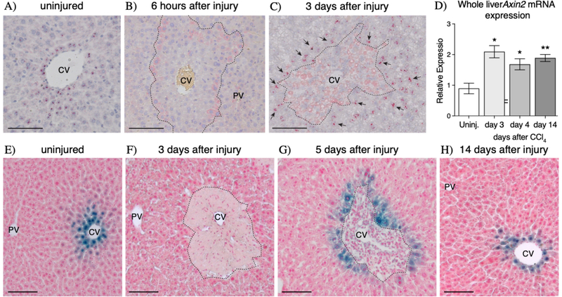
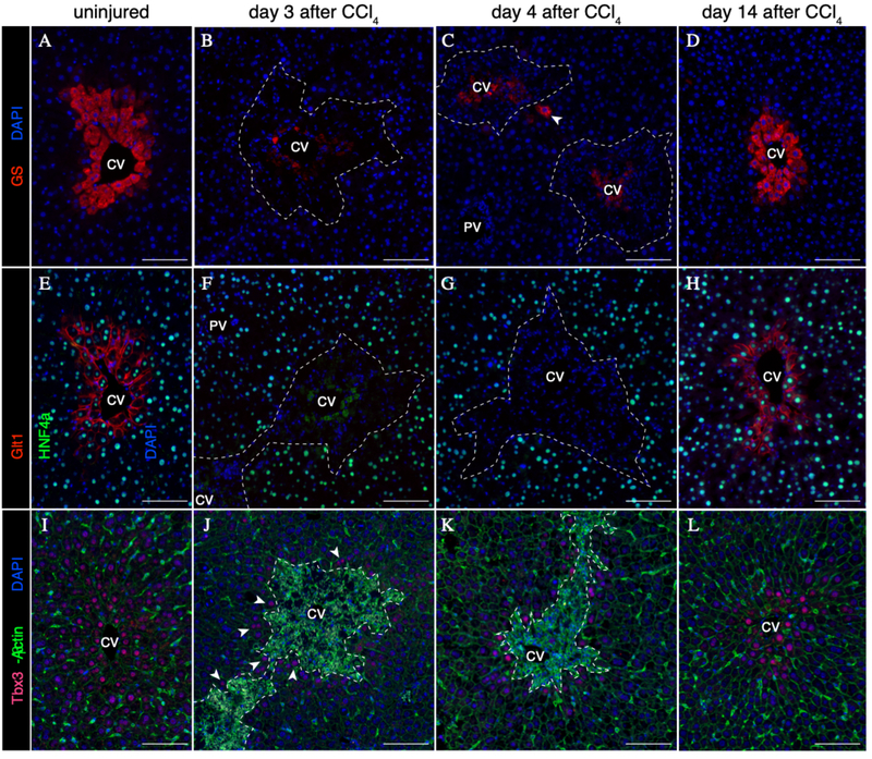
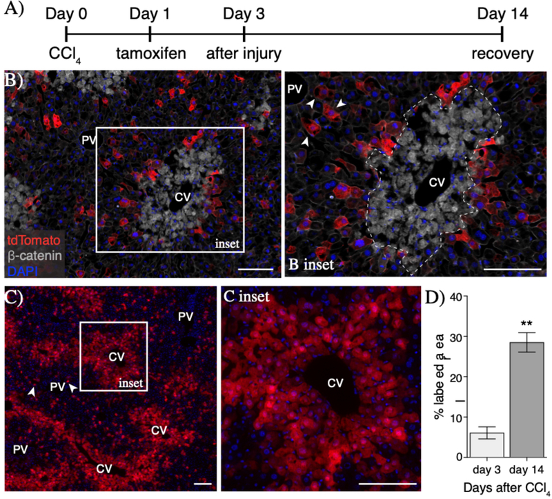
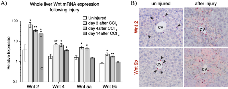
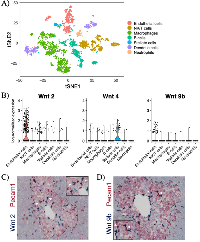
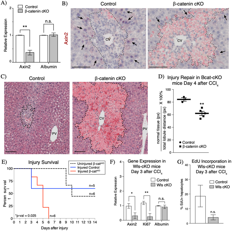
In the liver, Wnt/β-catenin signaling is involved in regulating zonation and hepatocyte proliferation during homeostasis. We examined Wnt gene expression and signaling after injury, and we show by in situ hybridization that Wnts are activated by acute carbon tetrachloride (CCl4 ) toxicity. Following injury, peri-injury hepatocytes become Wnt-responsive, expressing the Wnt target gene axis inhibition protein 2 (Axin2). Lineage tracing of peri-injury Axin2+ hepatocytes shows that during recovery the injured parenchyma becomes repopulated and repaired by Axin2+ descendants. Using single-cell RNA sequencing, we show that endothelial cells are the major source of Wnts following acute CCl4 toxicity. Induced loss of β-catenin in peri-injury hepatocytes results in delayed repair and ultimately injury-induced lethality, while loss of Wnt production from endothelial cells leads to a delay in the proliferative response after injury. Conclusion: Our findings highlight the importance of the Wnt/β-catenin signaling pathway in restoring tissue integrity following acute liver toxicity and establish a role of endothelial cells as an important Wnt-producing regulator of liver tissue repair following localized liver injury.


Figures

Figure 1
Figure 1
CCI4 injury induces de novo Axin2 expression in peri-injury hepatocytes. (A-C) Axin2 mRNA in situ hybridization in uninjured (A), early injured, and repairing livers show that Axin2 expression is lost as soon as 6 hours after injury (B) but is re-activated in peri-injury hepatocytes during injury repair (C, arrows). (D) Upregulation of Axin2 expression following injury is confirmed by whole liver qRT-PCR. (E-H) Axin2-LacZ reporter staining also confirms the dynamics of de novo Axin2 expression in peri-injury hepatocytes following CCl4 toxicity. 100μm scale bars shown. CV = central vein, PV = portal vein. Dashed line represents injury border. Error bars show S.E.M. *p<0.05, **p<0.01 by two-tailed t-test compared to uninjured control for n=3 animals per timepoint.
Figure 2
Figure 2
Post-CCl4 peri-injury hepatocytes do not express Wnt targets GS and Glt1, but are positive for Tbx3. (A-D) Expression of GS in uninjured (A) and post-injury (B-D) liver. Rare GS+ hepatocytes are observed during injury repair (C, arrowhead). (E-H) Expression of Glt1 in uninjured (E) and post-injury (B-D) liver. (I-L) Expression of Tbx3 in uninjured (I) and post-injury (J-L) liver. Low levels of Tbx3 expression are induced in peri-injury hepatocytes as early as 3 days following injury (J, arrowheads). 100μm scale bars. Dashed line represents injury border. CV = central vein, PV = portal vein. Representative images from n=3 animals per timepoint.
Figure 3
Figure 3
Peri-injury Axin2+ hepatocytes proliferate to repair damaged tissue. (A) Injury and lineage labeling scheme for panels B-D. Peri-injury Axin2+ hepatocytes are efficiently labeled following injury (B) and show significant expansion over the course of tissue repair (C). Notably, tdTomato+ hepatocytes in the periportal space (B inset, arrowheads) show no expansion upon injury repair (C, arrowheads). (D) Quantification of percent labeled area. 100μm scale bars. CV = central vein, PV = portal vein. Dashed line represents injury border. Error bars show S.E.M. **p<0.01 by two-tailed t-test for n=3 animals per timepoint.
Figure 4
Figure 4
Upregulation and expression pattern of Wnt ligands after injury. (A) Whole liver qRT-PCR results showed significant (p-val<0.05) upregulation of four Wnt ligands whose expression is found in the pericentral and mid-lobular regions following injury. (B) mRNA in situ hybridization for Wnt2 and Wnt9b show expanded regions of expression for these two pericentral Wnt ligands early after injury. Arrowheads indicate positive signal in the uninjured liver. In situ hybridization images for Wnt4 and Wnt5a can be found in Supplemental Figure 4. Dashed lines represent injury border. Error bars show S.E.M. *p<0.05, **p<0.01 by two-tailed t-test for n=3 animals per timepoint.
Figure 5
Figure 5
Endothelial cells are the major source of Wnt2 and Wnt9b after injury. (A) T-distributed stochastic neighbor embedding (tSNE) plot showing seven clusters of non-parenchymal cells in the day 3 CCl4-injured (n=3115 cells) livers. Colors denote different cell types as shown in the legend. Markers used to identify specific cell types are shown in Supplemental Figure 5D. (B) Violin plots of log-normalized expression (natural logarithm of 1+counts per 10,000) of Wnt2, Wnt4, and Wnt9b in the injured liver, categorized by cell type. Double in situ hybridization of Pecam1 (red) and (C) Wnt2 (blue) and (D) Wnt9b (blue) shows significant overlap between Pecam1 these two Wnts. Arrowheads denote examples of double-positive cells. Dashed line denotes injury border. N=3 animals for all studies shown.
Figure 6
Figure 6
Wnt/β-catenin signaling is necessary for injury repair and survival. (A) Inducible knockout of β-catenin in the CCl4 injured liver results in significant decrease in Axin2 expression as measured by whole liver qRT-PCR. (B) Axin2 expression is specifically downregulated in peri-injury hepatocytes (arrows) upon loss of β-catenin in injured Bcat-cKO mouse livers (n=3 control mice, n=5 Bcat-cKO mice). (C) H&E staining morphology of injured control and Bcat-cKO livers at 4 days after injury. (D) Quantification of the extent of parenchymal repopulation based on image analysis of H&E stained sections of injured livers (see Methods) show that injured B-cat cKO livers are significantly less repaired at 4 days following injury than compared to injured control livers. (E) Loss of β-catenin ultimately results in lethality following CCl4 injury. (F) Loss of Wntless in endothelial cells results in decreased Axin2 and Ki67 expression by whole liver qRT-PCR at 3 days after injury. (G) A concurrent decrease in hepatocyte EdU incorporation is also observed in Wls-cKO animals (n=4 control mice, n=3 Wls cKO mice). Error bars show S.E.M. *p<0.05, **p<0.01 by two tailed t-test, n.s. = not statistically significant. 100μm scale bars. CV = central vein, PV = portal vein. Dashed line represents injury border.

References

    1. Malato Y, Naqvi S, Schurmann N, Ng R, Wang B, Zape J, et al. Fate tracing of mature hepatocytes in mouse liver homeostasis and regeneration. J. Clin. Invest. 2011;121:4850–4860. - PMC - PubMed
    1. Yanger K, Knigin D, Zong Y, Maggs L, Gu G, Akiyama H, et al. Adult Hepatocytes Are Generatedby Self-DuplicationRather than Stem Cell Differentiation. Stem Cell. 2014;:1–22. - PMC - PubMed
    1. Newsome PN, Hussain MA, Theise ND. Hepatic oval cells: helping redefine a paradigm in stem cell biology. Curr. Top. Dev. Biol. 2004;61:1–28. - PubMed
    1. Huch M, Dorrell C, Boj SF, Van Es JH, Li VSW, van de Wetering M, et al. In vitro expansion of single Lgr5+ liver stem cells induced by Wnt-driven regeneration. Nature. 2013;494:247–250. - PMC - PubMed
    1. Tanimizu N, Mitaka T. Re-evaluation of liver stem/progenitor cells. Organogenesis. 2014;10:208–215. - PMC - PubMed

Publication types

MeSH terms