Review
. 2019 Mar;33(2):383-402.
doi: 10.1111/jvim.15406. Epub 2019 Feb 14.

Comparative pathophysiology and management of protein-losing enteropathy

Affiliations
Review

Comparative pathophysiology and management of protein-losing enteropathy

Melanie D Craven et al. J Vet Intern Med. 2019 Mar.

Abstract

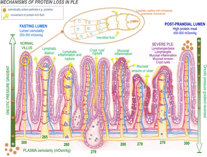
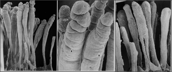
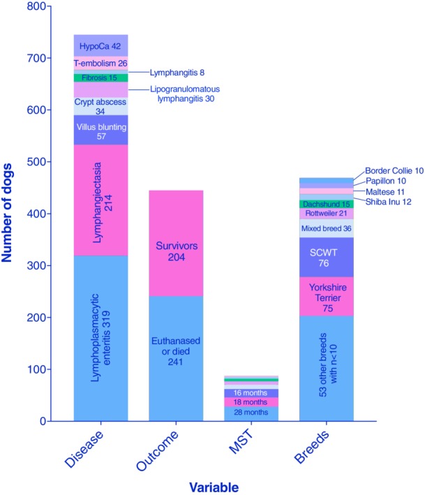
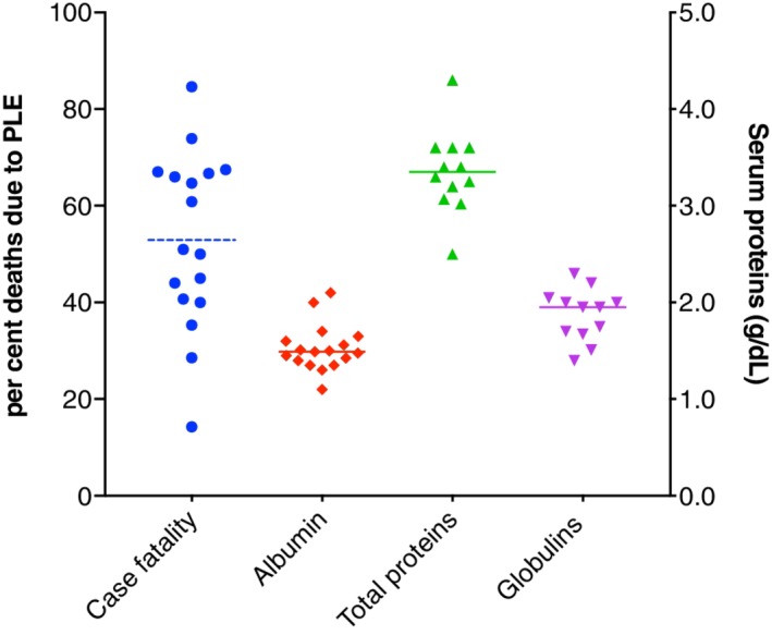
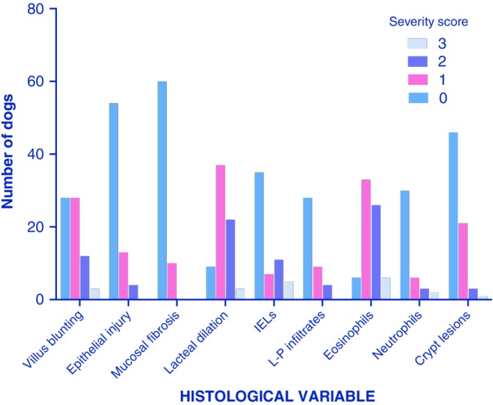
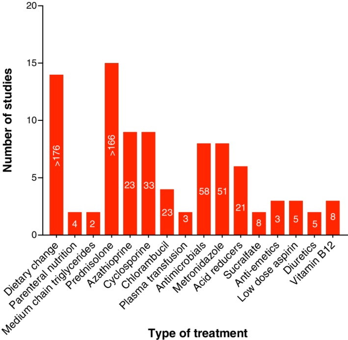
Protein-losing enteropathy, or PLE, is not a disease but a syndrome that develops in numerous disease states of differing etiologies and often involving the lymphatic system, such as lymphangiectasia and lymphangitis in dogs. The pathophysiology of lymphatic disease is incompletely understood, and the disease is challenging to manage. Understanding of PLE mechanisms requires knowledge of lymphatic system structure and function, which are reviewed here. The mechanisms of enteric protein loss in PLE are identical in dogs and people, irrespective of the underlying cause. In people, PLE is usually associated with primary intestinal lymphangiectasia, suspected to arise from genetic susceptibility, or "idiopathic" lymphatic vascular obstruction. In dogs, PLE is most often a feature of inflammatory bowel disease (IBD), and less frequently intestinal lymphangiectasia, although it is not proven which process is the true driving defect. In cats, PLE is relatively rare. Review of the veterinary literature (1977-2018) reveals that PLE was life-ending in 54.2% of dogs compared to published disease-associated deaths in IBD of <20%, implying that PLE is not merely a continuum of IBD spectrum pathophysiology. In people, diet is the cornerstone of management, whereas dogs are often treated with immunosuppression for causes of PLE including lymphangiectasia, lymphangitis, and crypt disease. Currently, however, there is no scientific, extrapolated, or evidence-based support for an autoimmune or immune-mediated mechanism. Moreover, people with PLE have disease-associated loss of immune function, including lymphopenia, severe CD4+ T-cell depletion, and negative vaccinal titers. Comparison of PLE in people and dogs is undertaken here, and theories in treatment of PLE are presented.

Keywords: PLE; crypt; hypoalbuminemia; lymphangiectasia; lymphatic; panhypoproteinemia.


Conflict of interest statement

Authors declare no conflict of interest.

Figures

Figure 1
Figure 1
Diagrammatic representation of “PLE syndrome” diseases in dogs (lymphangiectasia, lymphangitis, crypt lesions, IBD, mucosal ulceration) and the effect of Starling's forces on intestinal osmolality and the interstitial‐lumenal oncotic gradient. Protein leaks into the small intestine passively, via an oncotic gradient from the interstitium to the lumen. If osmotic pressures equilibrate, a reduction in passive leak is expected. When the intestinal lumen has a higher oncotic pressure than the interstitium, such as postprandially, the gradient may be reversed, depending on the underlying pathology. Top: Magnification of lymphatic capillary structure, showing overlapping endothelial cells that act as flap‐like doors allowing interstitial fluid to enter while preventing lymph exit. IBD, inflammatory bowel disease; PLE, protein‐losing enteropathy
Figure 2
Figure 2
A scanning electron microscopy view (left) of a corrosion cast of central lacteals in canine jejunum. A higher magnification (center) showing circular constrictions (arrow) near the top of the lacteals. Arrowheads show imprints of endothelial nuclei. Central lacteals in the ileum (right) are slender leaf‐like forms. ×110. From Yamanaka et al.,23 with permission
Figure 3
Figure 3
Cumulative data from 23 PLE articles spanning from 1977 to 2017, involving a total of 469 dogs. HypoCa, hypocalcemia; MST, median survival time; PLE, protein‐losing enteropathy; SCWT, Soft Coated Wheaten Terrier; T‐embolism, thromboembolism12, 13, 49, 50, 51, 52, 53, 54, 55, 56, 57, 58, 59, 60, 61, 62, 63, 64, 65, 66, 67, 68, 69
Figure 4
Figure 4
Breed frequencies represented across canine PLE publications; numbers on bars indicate the number of dogs within the breed.12, 13, 49, 50, 51, 52, 53, 54, 55, 56, 57, 58, 59, 60, 61, 62, 63, 64, 65, 66, 67, 68, 69 PLE, protein‐losing enteropathy
Figure 5
Figure 5
Cumulative disease‐associated deaths and serum proteins reported in PLE publications (solid line median, dotted line mean).12, 13, 49, 50, 51, 52, 53, 54, 55, 56, 57, 61, 64, 65, 66, 67, 69 PLE, protein‐losing enteropathy
Figure 6
Figure 6
Cumulative histology scores from 3 canine studies of PLE, per the WSAVA GI standardization group scoring system.48, 53, 65GI, gastrointestinal; PLE, protein‐losing enteropathy. Numbers on bars are the number of dogs
Figure 7
Figure 7
Cumulative treatment data in PLE across studies; white numbers on each bar indicate the minimum number of dogs that received each specific treatment.49, 51, 52, 53, 54, 55, 56, 57, 61, 64, 65, 66, 67, 69 PLE, protein‐losing enteropathy
Figure 8
Figure 8
Histology of endoscopic mucosal biopsies showing small intestinal crypt lesions from a Yorkshire Terrier with PLE (H&E stain; Aperio Digital Scan, 10×, left). Florescence in situ hybridization microscopic analysis (right, magnification ×200) with eubacterial probe (red), non‐eubacterial (green), and DAPI staining nuclear structures (blue) showing no evidence of intralesional or mucosally adherent bacteria.81 PLE, protein‐losing enteropathy

Similar articles

Cited by

References

    1. Braamskamp MJAM, Dolman KM, Tabbers MM. Clinical practice. Protein‐losing enteropathy in children. Eur J Pediatr. 2010;169(10):1179‐1185. - PMC - PubMed
    1. Freeman HJ, Nimmo M. Intestinal lymphangiectasia in adults. World J Gastrointest Oncol. 2011;3(2):19‐23. - PMC - PubMed
    1. Rovenská E, Rovenský J. Lymphatic vessels: structure and function. Isr Med Assoc J. 2011;13(12):762‐768. - PubMed
    1. Waldmann TA, Steinfeld JL, Dutcher TF, Davidson JD, Gordon RS. The role of the gastrointestinal system in idiopathic hypoproteinemia. Gastroenterology. 1961;41:197‐207. - PubMed
    1. Takeda H, Ishihama K, Fukui T, et al. Significance of rapid turnover proteins in protein‐losing gastroenteropathy. Hepatogastroenterology. 2003;50(54):1963‐1965. - PubMed

MeSH terms