

Comparison of postpartum sonographic findings after uneventful vaginal and cesarean section deliveries
- PMID: 30763015
- PMCID: PMC6444312
- DOI: 10.15557/JoU.2018.0045
Comparison of postpartum sonographic findings after uneventful vaginal and cesarean section deliveries
Abstract
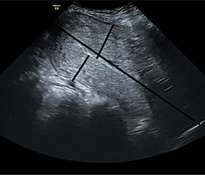
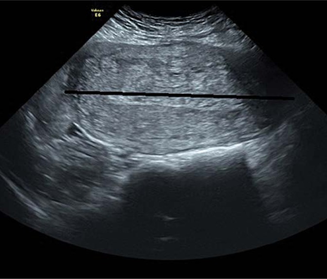
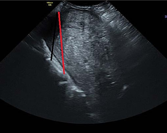
Objective: To prospectively determine the sonographic findings of the postpartum uterus 24 hours after vaginal delivery and cesarean section. Methods: Women who had uneventful vaginal delivery or cesarean section from July 2015 to May 2018 in a tertiary care hospital were prospectively included. Uterine lengths, endometrium, amout of free fluid, the distance between the uterine fundus-promontorium and uterine fundus-L5 were evaluated 24 hours after delivery. Results: The mean (min-max) endometrial thickness in the vaginal delivery and cesarean section groups were 13.3 (4-25) and 12.4 (4-29) mm, respectively. Fundus-cervix length was significantly higher in the vaginal delivery group compared to the cesarean section group (184.05 ± 16.8 vs 163.6 ± 6.7 mm, p <0.001). The measurements of anterior and anterior-posterior walls of the uterus, anteroposterior uterine length and uterine width were similar in both groups. Promontorium-fundus length was significantly higher in patients who delivered vaginally than those by cesarean section (123.3 ± 13.6 vs 108.7 ± 23.3 mm, p = 0.005). Conclusion: The measurement of L5-fundus distance is a simple and effective technique to evaluate the size of the uterus. Homogenous endometrium up to 30 mm in asymptomatic patients may be a normal finding 24 hours after delivery. The results of this study may be helpful in the decision-making process in cases of early postpartum hemorrhage or hemodynamic instability.
Objective: To prospectively determine the sonographic findings of the postpartum uterus 24 hours after vaginal delivery and cesarean section. Methods: Women who had uneventful vaginal delivery or cesarean section from July 2015 to May 2018 in a tertiary care hospital were prospectively included. Uterine lengths, endometrium, amout of free fluid, the distance between the uterine fundus-promontorium and uterine fundus-L5 were evaluated 24 hours after delivery. Results: The mean (min–max) endometrial thickness in the vaginal delivery and cesarean section groups were 13.3 (4–25) and 12.4 (4–29) mm, respectively. Fundus-cervix length was significantly higher in the vaginal delivery group compared to the cesarean section group (184.05 ± 16.8 vs 163.6 ± 6.7 mm, p <0.001). The measurements of anterior and anterior-posterior walls of the uterus, anteroposterior uterine length and uterine width were similar in both groups. Promontorium-fundus length was significantly higher in patients who delivered vaginally than those by cesarean section (123.3 ± 13.6 vs 108.7 ± 23.3 mm, p = 0.005). Conclusion: The measurement of L5-fundus distance is a simple and effective technique to evaluate the size of the uterus. Homogenous endometrium up to 30 mm in asymptomatic patients may be a normal finding 24 hours after delivery. The results of this study may be helpful in the decision-making process in cases of early postpartum hemorrhage or hemodynamic instability.
Figures
Similar articles
-
Sonographic appearance of the uterus in the early puerperium in vaginal versus cesarean deliveries: a prospective study.J Matern Fetal Neonatal Med. 2018 Aug;31(15):1983-1988. doi: 10.1080/14767058.2017.1333099. Epub 2017 Jun 2. J Matern Fetal Neonatal Med. 2018. PMID: 28521590
-
Abdominal and pelvic ultrasound findings within 24 hours following uneventful Cesarean section.Ultrasound Obstet Gynecol. 2008 Sep;32(4):520-6. doi: 10.1002/uog.6120. Ultrasound Obstet Gynecol. 2008. PMID: 18683208
-
Changes in uterine size after vaginal delivery and cesarean section determined by vaginal sonography in the puerperium.Arch Gynecol Obstet. 1999 Nov;263(1-2):13-6. doi: 10.1007/s004040050253. Arch Gynecol Obstet. 1999. PMID: 10728621
-
[Uterine scar after caeserean section- predicting the risk of uterine rupture and decision on the way of delivery].Akush Ginekol (Sofiia). 2014;53(4):29-32. Akush Ginekol (Sofiia). 2014. PMID: 25510068 Review. Bulgarian.
-
Delivery for women with a previous cesarean: guidelines for clinical practice from the French College of Gynecologists and Obstetricians (CNGOF).Eur J Obstet Gynecol Reprod Biol. 2013 Sep;170(1):25-32. doi: 10.1016/j.ejogrb.2013.05.015. Epub 2013 Jun 28. Eur J Obstet Gynecol Reprod Biol. 2013. PMID: 23810846 Review.
Cited by
-
Preliminary study of the effect of low-intensity focused ultrasound on postpartum uterine involution and breast pain in puerperal women: a randomised controlled trial.Sci Rep. 2024 Jan 5;14(1):658. doi: 10.1038/s41598-024-51328-9. Sci Rep. 2024. PMID: 38182657 Free PMC article. Clinical Trial.
-
The Importance of the Novel Postpartum Uterine Ultrasonographic Scale in Numerical Assessments of Uterine Involution Regarding Perinatal Maternal and Fetal Outcomes.Diagnostics (Basel). 2021 Sep 21;11(9):1731. doi: 10.3390/diagnostics11091731. Diagnostics (Basel). 2021. PMID: 34574072 Free PMC article.
References
-
- Al-Zirqi I, Vangen S, Forsen L, Stray-Pedersen B: Prevalence and risk factors of severe obstetric haemorrhage. BJOG 2008; 115: 1265–1272. - PubMed
-
- Plunk M, Lee JH, Kani K, Dighe M: Imaging of postpartum complications: A multimo-dality review. AJR Am J Roentgenol 2013; 200: W143–W154. - PubMed
-
- Thomassin-Naggara I, Darai E, Bazot M: Gynecological pelvic infection: What is the role of imaging? Diagn Interv Imaging 2012; 93: 491–499. - PubMed
-
- Kamaya A, Ro K, Benedetti NJ, Chang PL, Desser TS: Imaging and diagnosis of postpartum complications: Sonography and other imaging modalities. Ultrasound Q 2009; 25: 151–162. - PubMed
-
- Luo A, Mao P: Late postpartum hemorrhage due to placental and fetal membrane residuals: Experience of two cases. Clin Exp Obstet Gynecol 2015; 42: 104–105. - PubMed
LinkOut - more resources
Full Text Sources