. 2019 Mar 26;13(3):3151-3161.
doi: 10.1021/acsnano.8b08668. Epub 2019 Feb 25.

Particle-Attachment-Mediated and Matrix/Lattice-Guided Enamel Apatite Crystal Growth

Affiliations

Particle-Attachment-Mediated and Matrix/Lattice-Guided Enamel Apatite Crystal Growth

Jacob R Jokisaari et al. ACS Nano. .

Abstract

Tooth enamel is a hard yet resilient biomaterial that derives its unique mechanical properties from decussating bundles of apatite crystals. To understand enamel crystal nucleation and growth at a nanoscale level and to minimize preparation artifacts, the developing mouse enamel matrix was imaged in situ using graphene liquid cells and atomic resolution scanning transmission electron and cryo-fracture electron microscopy. We report that 1-2 nm diameter mineral precipitates aggregated to form larger 5 nm particle assemblies within ameloblast secretory vesicles or annular organic matrix subunits. Further evidence for the fusion of 1-2 nm mineral precipitates into 5 nm mineral aggregates via particle attachment was provided by matrix-mediated calcium phosphate crystal growth studies. As a next step, aggregated particles organized into rows of 3-10 subunits and developed lattice suprastructures with 0.34 nm gridline spacings corresponding to the (002) planes of apatite crystals. Mineral lattice suprastructures superseded closely matched organic matrix patterns, suggestive of a combination of organic/inorganic templates guiding apatite crystal growth. Upon assembly of 2-5 nm subunits into crystal ribbons, lattice fringes indicative of the presence of larger ordered crystallites were observed surrounding elongating crystal ribbons, presumably guiding the c-axis growth of composite apatite crystals. Cryo-fracture micrographs revealed reticular networks of an organic matrix on the surface of elongating enamel crystal ribbons, suggesting that protein coats facilitate c-axis apatite crystal growth. Together, these data demonstrate (i) the involvement of particle attachment in enamel crystal nucleation, (ii) a combination of matrix- and lattice-guided crystal growth, and (iii) fusion of individual crystals via a mechanism similar to Ostwald ripening.

Keywords: apatite; atomic scale microscopy; crystal growth; enamel; graphene liquid cell.


Figures
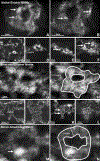
Figure 1.
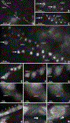
Figure 1.. Enamel matrix subunit compartments from native enamel matrix, sonicated enamel matrix and in ameloblast secretory vesicles imaged using aberration corrected STEM imaging.
(A,B) Crystal nucleation in organic subunit compartments within the native enamel matrix of 1 days postnatal developing mouse molars. Note the individual 1–3nm diameter nucleation sites (arrows, A,B) randomly dispersed with a circular 20nm diameter organic matrix assembly (matrix)(A,B). (C-F) Network of 20nm subunit compartments generated from the sonicated 3dpn mouse molar enamel layer and its adjacent enamel matrix (matrix). Note the dense mineral assemblies (arrows) associated with protein subunits. The delineated square outlines the area selected for the individual subunit magnification in Figs. (G,H). (G,H) Nanoscale resolution image of the 20nm diameter organic matrix subunits from the sonicated enamel matrix. (I-N) Ultra high-resolution micrographs of the enamel matrix immediately adjacent to the secretory ameloblast layer. Note the presence of isolated mineral condensations within each protein subunit compartment (arrows). The rectangle demarks the area used for the high magnification micrographs in Figs. M,N. Nanoscale resolution image of the 20nm diameter organic matrix subunits from the ameloblast secretory vesicles (M,N). The site of mineral condensation (min) was restricted to one corner of the organic matrix (matrix) subunit compartment (N).
Figure 2.
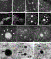
Figure 2.. Particle-attachment mediated crystal growth and lattice-guided elongation in the native enamel matrix of 1 day postnatal developing mouse molars via atomic resolution STEM.
(A-D) Linear rows of longitudinally aligned individual nucleation sites (arrows) surrounded by organic matrix (matrix). Note the regular pattern of 0.3–0.4nm distance parallel lattice fringes perpendicular to the direction of individual nucleation sites indicative of the presence of crystallized matter. (A) Particle attachment stage, demonstrating three nucleation sites (arrows) surrounded by organic matrix (matrix), with individual particles aggregated in the proximity of each nucleation site. (B) Three rows of 5–7 linear arranged nucleation sites surrounded by organic matrix and connected through lattice fringes, each measuring approximately 20nm in length. (C,D) Lattice guided bridging of mineralization sites (arrows). Individual nucleation sites were 4–8nm apart from each other, and lattice fringes extended beyond nucleation sites. (E-J) Relationship between widened crystalline lattice structures (lattice) and matrix interfaces (matrix) during continued enamel apatite crystal growth. (E-G) High magnification STEM micrographs of three adjacent 3nm diameter crystals surrounded by organic matrix (matrix). Parallel lattice fringes at a 0.3–0.4nm distance from each other were indicative of the presence of newly formed apatite crystals within the enamel matrix. (H-J) High magnification focus stack of an initial apatite crystal nucleation site within the organic matrix (matrix), revealing parallel 0.3–0.4nm distance lattice fringes and grid patterns. (K-M) High resolution ultrastructure of an elongated composite enamel crystal. Note the composite structure of this initial enamel crystal needle consisting of individual crystalline subunits (a-f). (L) Criss-cross lattice structure in adjacent crystal subunits. Note the bright lattice fringes parallel to the shaft (shaft) of the crystal needle (double arrows, H and I). (M) Lattice organization of the crystalline subunit at the enamel crystal needle tip. Note the distinct lattice fringes parallel to the elongated crystal needle (double arrows). Protein matrix patterns (matrix) were detected in proximity to the tip of the elongated crystal.
Figure 3.
Figure 3.. Enamel crystal growth studies within the STEM ARM liquid cell in vitro.
(A-D) Mineral deposition within the 20nm diameter organic matrix subunits after 1 hour incubation. Mineral precipitates are indicated with an arrow (A-D). (E-N) Mineral deposition and crystal nucleation within the 20nm diameter organic matrix subunits after 2 hours incubation. A-H were taken in high angle annular dark field mode, I-L in low angle annular dark field mode, and M-P in annular bright field mode. The position of the protein matrix (matrix), of the nucleating apatite crystals (HA) is indicated, and of the lattice spacings of newly formed crystals (lattice) is marked. Arrowheads (H, J) indicate the position of the protein- and mineral- free zone surrounding newly formed crystallites.
Figure 4.
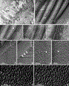
Figure 4.. Cryo-EM analysis of freeze-fractured 3 days postnatal developing tooth enamel and enamel organs.
(A) Overview micrograph illustrating the freeze-fracture topography at the interface between ameloblasts (amel), (dentin) (de) and predentin (pd). There was a thin layer of protein matrix (ma) between the ameloblast cell layer (amel) and the dentin layer (de). (B-K) are freeze-fracture cryo electron micrographs from the early enamel layer positioned between ameloblasts (amel) and dentin (de). (B) Freeze fracture topography of the organic matrix (prot) immediately associated with the inorganic crystal surface (cryst). (C,D) Identification of 50–100nm annular protein matrix assembly rings (arrowheads) on crystal surfaces. (D,E) 20nm spherical matrix subunit position on the crystal surface (double arrows). (F-I) Protein matrix at the ameloblast face of developing enamel crystals (arrows, G). The arrowheads in (H, I) point to annular subunit compartments measuring approximately 50–100nm in diameter. (J,K) image processing technologies were applied to either enhance the contrast (J) or to emboss the 3D surface relief (K) of the micrograph in (H) to further define the structural basis of enamel protein assemblies on crystal surfaces.
Figure 5.
Figure 5.. Nanoscale structure of unfixed 3 days postnatal first mouse molar enamel crystals as observed via atomic resolution STEM (A-C) and freeze-fracture scanning microscopy (D,E).
(A) Overview micrograph demonstrating individual crystals (cryst) organized into enamel prisms (prism). The insert indicates the area chosen for higher magnification micrographs in (B) and (C). (A-C) individual enamel crystals. Note the irregularities along the crystal c-axis and the close proximity between subunits from adjacent crystals (cryst). (D) Cryofracture micrographs illustrating enamel crystal morphology (cryst) in vertical direction. Note the serrated lateral surface of the elongated enamel crystals (arrowheads). (E) SEM visualization of the cryo-etched enamel prisms (prism). Prisms containing individual crystals are highlighted.
Figure 6.
Figure 6.. (Sketch) Five successive stages of enamel crystal precipitation and elongation according to our STEM and cryo-fracture data.
(A) Stage of initial calcium phosphate particle precipitation and attachment within the organized enamel protein matrix, (B) Formation of initial crystal needles through lattice- and matrix-guided bridging of individual nucleation sites, (C) Lattice-guided alignment and extension of individual apatite crystals into elongated enamel crystals, (D) Recently fused early enamel crystal with surrounding lattices guiding further crystal elongation and growth, (E) Decussating arrangement of individual enamel crystals into enamel prisms (rods). The oblate ellipsoids on the surface of individual enamel crystals illustrate the shape of the protein coat based on our cryo-fracture micrographs.

Similar articles

Cited by

References

    1. Gopinathan G; Jin T; Liu M, Li S; Atsawasuwan P; Galang MT; Allen M; Luan X; Diekwisch TGH The Expanded Amelogenin Polyproline Region Preferentially Binds to Apatite versus Carbonate and Promotes Apatite Crystal Elongation. Front. Physiol 2014, 5, 430. - PMC - PubMed
    1. Lowenstam HA Minerals Formed by Organisms. Science 1981, 211, 1126–1131. - PubMed
    1. Mann S Biomineralization: Principles and Concepts in Bioinorganic Materials Chemistry Oxford University Press: Oxford, UK, 2001; pp 3.
    1. Diekwisch T; David S; Bringas P Jr; Santos V; Slavkin HC Antisense Inhibition of AMEL Translation Demonstrates Supramolecular Controls for Enamel HAP Crystal Growth during Embryonic Mouse Molar Development. Development 1993, 117, 471–482. - PubMed
    1. Diekwisch TG; Berman BJ; Gentner S; Slavkin HC Initial Enamel Crystals are not Spatially Associated with Mineralized Dentine. Cell Tissue Res 1995, 279, 49–67. - PubMed

Publication types