. 2018 Apr 1;157(4):383-398.
doi: 10.1530/REP-18-0386.

Cyclin-dependent kinase 21 is a novel regulator of proliferation and meiosis in the male germline of zebrafish

Affiliations

Cyclin-dependent kinase 21 is a novel regulator of proliferation and meiosis in the male germline of zebrafish

Kaitlyn A Webster et al. Reproduction. .

Abstract

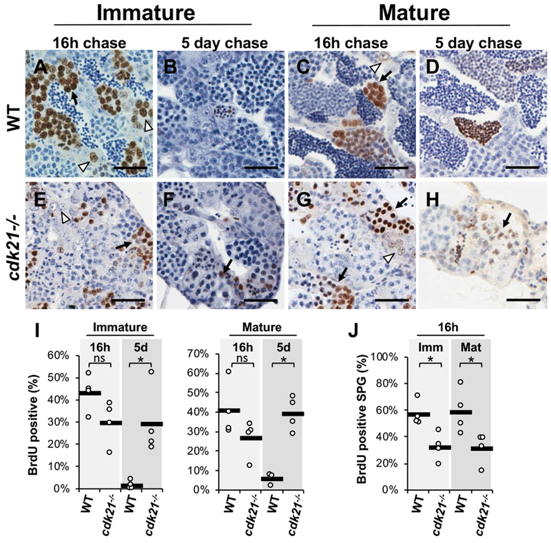
Germ cell differentiation and maintenance relies on complex regulation of mitotic and meiotic progression. Cyclin-dependent kinases (CDKs) and their activating cyclin partners are known to have specialized roles in regulating cell cycle progression across tissues, including germ cells. Very little is known about CDK/cyclin function in zebrafish or the regulation of germ cell maintenance and differentiation. In a forward genetic screen for gonadogenesis defects in zebrafish, a mutation disrupting cdk21 (cyclin-dependent kinase 21) was identified, which caused gonad hypoplasia, reduced fertility and failure of female sex specification. The cdk21 gene is unique to fishes, though the encoded protein is related to the D-cyclin partners Cdk4 and Cdk6, which are known G1 cell cycle regulators. In the testis, cdk21 mutant germ cells exhibited cell cycle defects such as diminished proliferation, prolonged meiosis and delayed sperm differentiation. Furthermore, cdk21 mutants failed to maintain germ cells following breeding. Based on these findings, we propose that cdk21 regulates spermatogonial proliferation, progression through meiosis and germline stem cell activation in the testis. In addition, we investigated cdk4 and cdk6 in zebrafish development and found that each has distinct expression patterns in the gonads. Mutant analysis demonstrated that cdk6 was necessary for viability beyond larval stages. In contrast, cdk4 mutants were viable but were all male with low breeding success and sperm overabundance. Our analysis demonstrated that zebrafish harbor three genes of the cdk4/6 family, cdk4, cdk6 and cdk21, with cdk21 having an essential role in germ cell development in the testis.


Conflict of interest statement

Declaration of Interest

The authors have no conflict of interests to declare.

Figures

Figure 1:
Figure 1:
cdk21 is a male germ cell enriched G1 cyclin-dependent kinase in fish. A) Wild-type adult female zebrafish have rounded abdomen (A), little yellow fin pigmentation and an external genital papilla (arrow, A’). B) Wild-type adult male zebrafish are slender (B) with prominent yellow pigmentation, no external genital papilla (B’), and develop paired elongated testes (B”) which appear opaque when sperm is abundant. C) Adult cdk21−/− males have male pigmentation (C’) and clear, hypoplastic testes (C”). D) Unrooted maximum likelihood analysis of known zebrafish cell cycle regulating cyclin-dependent kinase proteins shows that Cdk21 clusters with Cdk4 and Cdk6. E) Cdk21 protein groups with Cdk4 and Cdk6 and was found broadly in bony fishes, but not in tetrapods. The chordate Ciona possess a single Cdk4/6 orthologue. F-I) Section in situ hybridization of adult zebrafish gonads. F, H) The vasa transcript indicates spermatogonia in the testes (arrowhead, F), and stages I and II oocytes in the ovary (H). G) The cdk21 transcript was expressed in the spermatogonia of the testes (arrowhead, G). I) In the ovary it was not detected in oogonia (lower inset) but was expressed at low levels in the cytoplasm of stage II oocytes as well as the Balbiani body (upper inset) of stage Ib oocytes. Oocyte staging according to Maack and Segner 2003. * = testes; arrow = genital papilla; sb = swim bladder; arrowhead = spermatogonia; og = oogonia; scale bars = 50 microns.
Figure 2:
Figure 2:
Loss-of-function mutations in the cdk21 gene cause spermatogenesis defects and preclusion of ovary fate. A-D, F-H) H&E staining of sections of adult zebrafish testes. A) Wild-type adult zebrafish testes contained discrete cysts of germ cells ranging from mitotic spermatogonia (white arrow) to meiotic spermatocytes (bracket), and lumens populated by sperm (black arrow). B-D) Adult homozygous cdk21t30421, cdk21umb7, and transheterozygous cdk21t30421/umb7 testes contained mitotic and meiotic germ cells but displayed little to no sperm. Insets show germ cells with abnormal nuclei. E) cdk21t30421 mutants were 100% male. WT includes cdk21+/+ and cdk21+/− (N=42). F) cdk21 t30421 testis following multiple successive breeding were nearly devoid of germ cells. G-H) Testes from one year old cdk21t30421 mutant (G) and wild-type sibling (H) maintained in the absence of females. I-J) H&E stain of juvenile gonads at 3 weeks post fertilization (wpf). I) 5 of 5 gonads from 3 wpf heterozygous fish contained oocytes and were classified as juvenile ovaries. J) 4 of 5 cdk21t30421 siblings contained only undifferentiated gonial cells within gonads and with no oocytes present. K) Testes from adult cdk21t30421 had increased amounts of cleaved Caspase-3 positive germ cells; horizontal bars denote the mean (P=0.017). White arrow = spermatogonia; bracket = spermatocytes; black arrow = sperm; asterisk = oocytes; WT = wild type. Scale bars = 20 μm.
Figure 3:
Figure 3:
The cdk21 gene is a regulator of proliferation and differentiation in male germ cells. A-H) Immunohistochemistry of BrdU positive cells in testes from wild-type male zebrafish and cdk21t30421 mutants exposed to BrdU for 18 hours followed by either 16 hours (16h) or 5 days (5d) of no BrdU exposure (chase). A, B) In immature (~2 mpf) wild-type testes, BrdU marked spermatogonia and primary spermatocytes after the 16h chase and spermatids after 5d chase. C, D) In mature (~4 mpf) wild-type testes, BrdU labeled spermatogonia and primary spermatocytes after 16h chase and spermatids/spermatozoa after 5d chase. E-H) Immature and mature cdk21 mutant testes had BrdU labeling in spermatogonia and spermatocytes after 16hr chase, and in primarily spermatocytes and spermatid/spermatozoa after 5d chase. I) Quantification of total BrdU positive cells (excluding post-meiotic cells). After 5d chase, in wild types very few BrdU labled cells were spermatogonia or spermatocytes. In mutant testes, many BrdU labeled cells had not yet progressed to spermiogenesis in mutants (P = 0.03, 5d immature; P = 0.03 5d mature). J) Quantification of BrdU labeled spermatogonia after 16h chase. The percentage of BrdU labeled spermatogonia out of total spermatogonia was calculated. Both immature and mature mutant testes had significantly fewer BrdU labeled spermatogonia (P = 0.03; immature; P = 0.03 mature). Horizontal black bars on all charts denote the mean. White arrowhead = spermatogonia; arrow = spermatocytes; WT = wild type; ns = not significant. Scale bar = 20 microns.
Figure 4:
Figure 4:
cdk21 mutant germ cells exhibit ectopic expression of Sycp3. A-P) Immunofluorescence on sections of adult testes. Wild-type (A-D) and cdk21t30421 mutant (E-H) testes labeled for α-Tubulin, DNA (DAPI), and Sycp3. Examples of germ cells cysts containing spermatogonia (white dashed), primary spermatocytes (turquoise dash-dotted), and spermatozoa (orange dotted) are outlined. I-P) High magnification of testicular germ cells showing localization of α-Tubulin (green), DNA (blue), and Sycp3 (magenta). I,M) Early-stage spermatogonia have relatively uncondensed DNA and prominent nucleoli (arrowhead). Wild-type spermatogonia (I) have little to no detectible Sycp3, whereas cdk21 mutant spermatogonia (M) frequently had cytoplasmic Sycp3 that often aggregated in puncta (arrow). Wild-type (J-K) and mutant (N-O) spermatocytes exhibited Sycp3 associated with chromosomes. Mutant sperm (P) displayed a pronounced Sycp3 compared to wild-type sperm (L). Q) Western blots of protein lysates extracted from zebrafish testes. Sycp3 was more abundant in mutant testes than wild-type. Tubulin was used as a loading control. Lepto-zygo = leptotene to zygotene stage spermatocytes. White arrowhead = nucleoli; white arrow = cytoplasmic Sycp3 puncta. Scale bar = 20 microns
Figure 5:
Figure 5:
The G1 cyclin-dependent kinases have distinct expression patters. A, B) RT-PCR from zebrafish tissues. A) mRNA expression of cdk4, cdk6, and cdk21 in tissues from adult fish. B) mRNAs expression in whole 24 hpf embryos, or torsos of 1 wpf, 2 wpf, and 3 wpf zebrafish. C-H) Section in situ hybridizations of adult testes and ovaries. C, F) cdk4 transcript was not detectable in the testes but localized prominently to the cytoplasm of stage Ib oocytes. D, G) cdk6 was detected uniformly throughout the testes, but in oocytes was restricted to the Balbiani body in stage Ib oocytes and the vegetal cortex of stage II oocytes. E) The rb1 transcript was expressed throughout the testes but was enriched in spermatogonia. H) In ovaries, rb1 transcript was localized to the cytoplasm of stages Ib oocytes. Oocyte staging according to Maack and Segner 2003. Scale bars = 20 microns
Figure 6:
Figure 6:
cdk4 and cdk6 mutant phenotypes do not resemble loss of cdk21. A,B,D) H&E stained sections of adult testes. A) Wild-type adult testes. B) cdk4umb8 mutants had histologically normal testes except for overabundance of sperm. C) Quantitation of the area occupied by sperm / the total area occupied by all germ cells (including sperm) from histological sections; the black bar shows the mean (N= 6 individuals per genotype, P = 0.002). D) cdk21t30421;cdk4umb8 double mutant testes resembles that of cdk21t30421 single mutants. E) Wild-type larva at 5 dpf showing an inflated swim bladder (arrow). F) cdk6umb9 at 5 dpf showing lack of swim bladder. F) cdk4umb8;cdk6umb9 double mutants displayed a range of severe developmental defects by 5 dpf, including short/wide body, lack of swim bladder, craniofacial deformities, and edemas. Scale bar = 20 microns

Similar articles

Cited by

References

    1. Anderson JL, Marí A, Braasch I, Amores A, Hohenlohe P, Batzel P and Postlethwait JH (2012) Multiple sex-associated regions and a putative sex chromosome in zebrafish revealed by RAD mapping and population genomics. PLoS ONE 7. - PMC - PubMed
    1. Ashley T, Walpita D and de Rooij DG (2001) Localization of two mammalian cyclin dependent kinases during mammalian meiosis. Journal of Cell Science 114 685–693. - PubMed
    1. Bowen ME, Henke K, Siegfried KR, Warman ML and Harris MP (2012) Efficient mapping and cloning of mutations in zebrafish by low-coverage whole-genome sequencing. Genetics 190 1017–1024. - PMC - PubMed
    1. Bradley KM, Breyer JP, Melville DB, Broman KW, Knapik EW and Smith JR (2011) An SNP-based linkage map for zebrafish reveals sex determination loci. Genes|Genomes|Genetics 1 3–9. - PMC - PubMed
    1. Daane JM, Rohner N, Konstantinidis P, Djuranovic S and Harris MP (2016) Parallelism and epistasis in skeletal evolution identified through use of phylogenomic mapping strategies. Molecular Biology and Evolution 33 162–173. - PMC - PubMed

Publication types