. 2019 Mar 21;4(6):e125326.
doi: 10.1172/jci.insight.125326.

PD-L1 on invasive fibroblasts drives fibrosis in a humanized model of idiopathic pulmonary fibrosis

Affiliations

PD-L1 on invasive fibroblasts drives fibrosis in a humanized model of idiopathic pulmonary fibrosis

Yan Geng et al. JCI Insight. .

Abstract

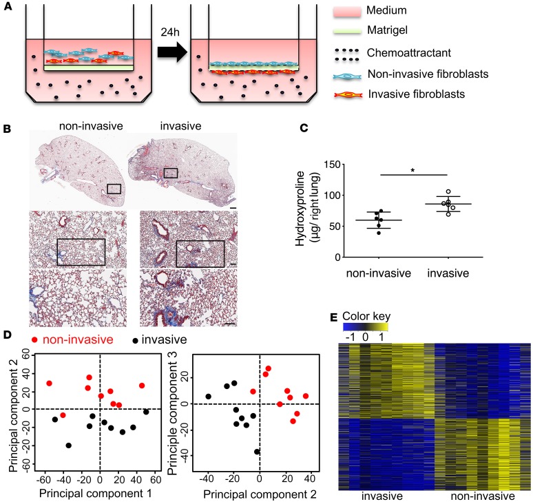
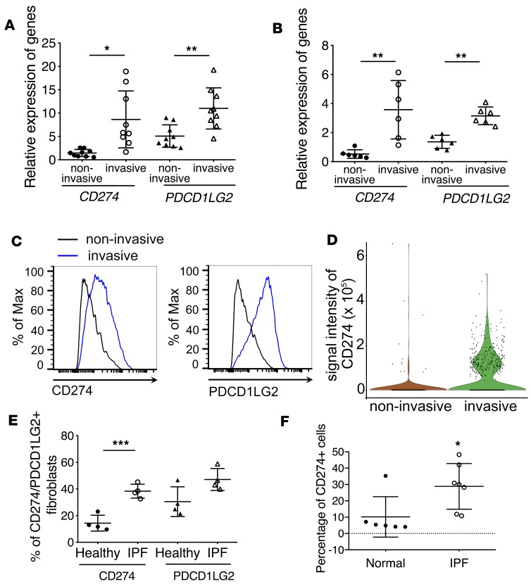
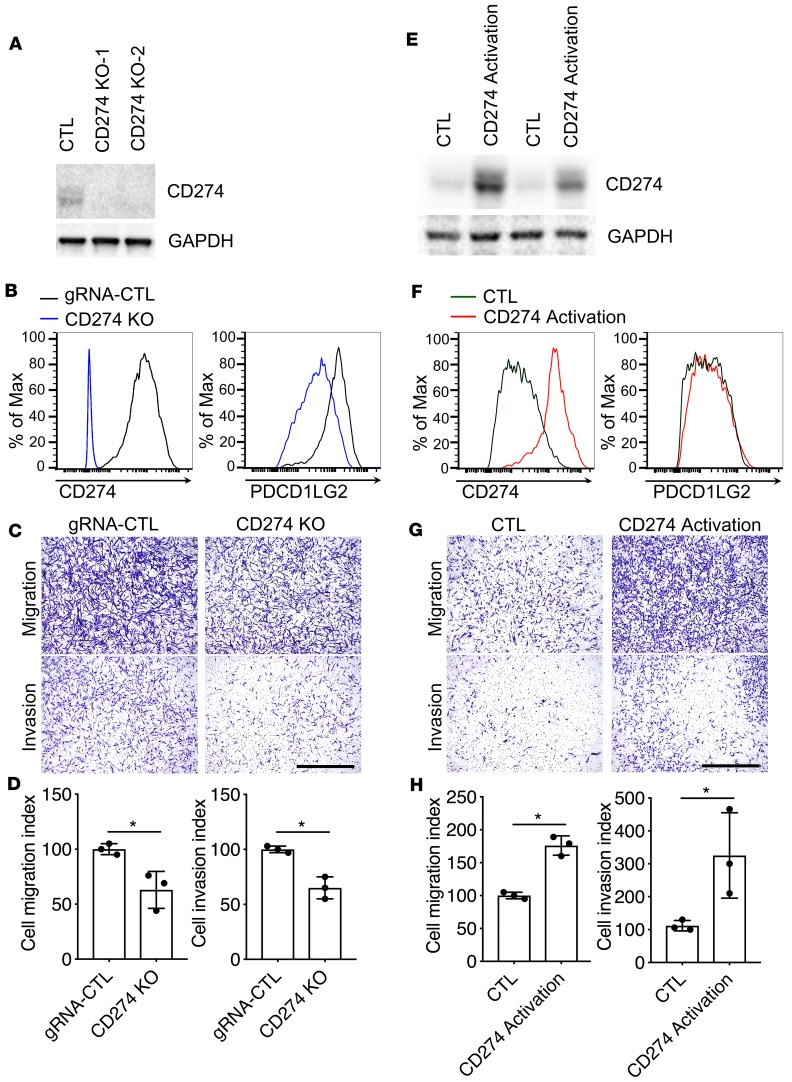
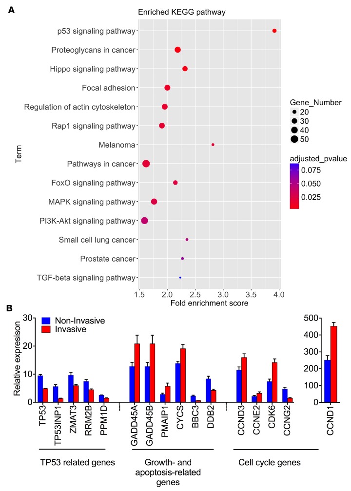
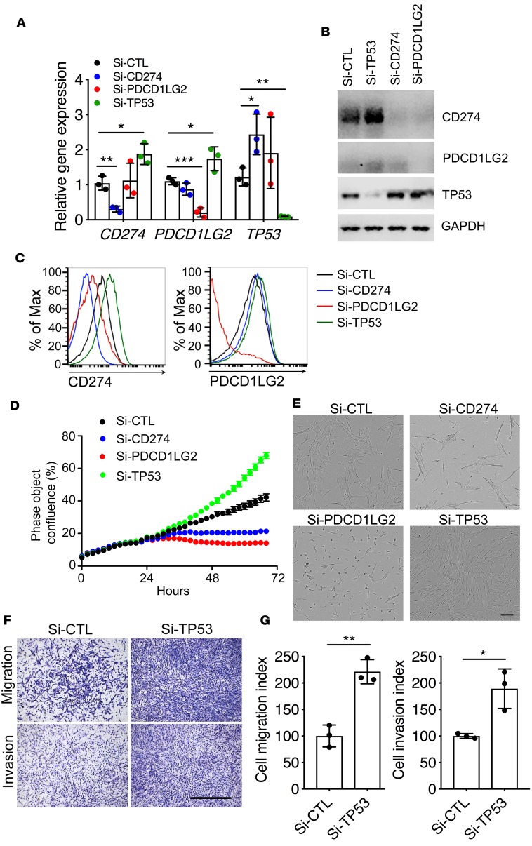
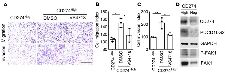
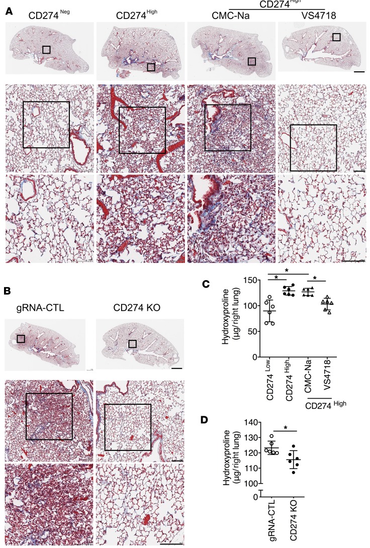
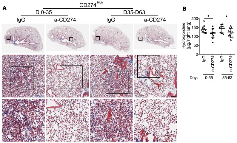
Idiopathic pulmonary fibrosis (IPF) is a progressive disease with unremitting extracellular matrix deposition, leading to a distortion of pulmonary architecture and impaired gas exchange. Fibroblasts from IPF patients acquire an invasive phenotype that is essential for progressive fibrosis. Here, we performed RNA sequencing analysis on invasive and noninvasive fibroblasts and found that the immune checkpoint ligand CD274 (also known as PD-L1) was upregulated on invasive lung fibroblasts and was required for the invasive phenotype of lung fibroblasts, is regulated by p53 and FAK, and drives lung fibrosis in a humanized IPF model in mice. Activating CD274 in IPF fibroblasts promoted invasion in vitro and pulmonary fibrosis in vivo. CD274 knockout in IPF fibroblasts and targeting CD274 by FAK inhibition or CD274-neutralizing antibodies blunted invasion and attenuated fibrosis, suggesting that CD274 may be a novel therapeutic target in IPF.

Keywords: Cell migration/adhesion; Fibrosis; Pulmonology; Respiration; Therapeutics.


Conflict of interest statement

Conflict of interest: The authors have declared that no conflict of interest exists.

Figures

Figure 1
Figure 1. Invasive lung fibroblasts promoted interstitial lung fibrosis.
(A) Schematic representation of in vitro invasion assay. Lung fibroblasts were seeded in the upper part of transwells. Cells attached to the bottom of Matrigel-coated membrane after 24 hours were considered invasive fibroblasts. Cells remaining on top of the Matrigel-coated membrane were considered noninvasive fibroblasts. Invasive and noninvasive IPF lung fibroblasts (n = 9 per group) were isolated using the matrigel invasion assay. Masson’s trichrome staining of collagen on lung sections (B) and hydroxyproline content in lung tissues (C) from NSG mice 50 days after injection with invasive and noninvasive IPF lung fibroblasts on day 50 after fibroblast injection (n = 6 per group). Scale bars: 1 mm (top panel), 100 μm (middle and lower panels). (D) Principal component analysis of RNA-seq data. (E) Heatmap of all differentially expressed (DE) genes in RNA-seq data. A total of 1,405 DE genes were identified with FDR < 0.01 and |log2 FC| > 0.5; among them, 719 DE genes were upregulated, and 686 DE genes were downregulated. *P < 0.05 by Student’s t test (C).
Figure 2
Figure 2. Upregulation of PD-1 ligands in invasive fibroblasts.
(A and B) Upregulation of immune checkpoint CD274 and PDCD1LG2 in invasive lung fibroblasts. RNA-seq (n = 9 per group) (A) and qRT-PCR analysis (n = 6 per group) (B) of CD274 and PDCD1LG2 expression in invasive and noninvasive IPF lung fibroblasts. (C) Cell surface expression of CD274 and PDCD1LG2 in invasive and noninvasive IPF lung fibroblasts. (D) Single-cell Western blot analysis of CD274 expression in invasive and noninvasive lung fibroblasts. (E) Cell surface expression of CD274 and PDCD1LG2 in primary IPF fibroblasts and healthy controls by flow cytometry. (F) Flow cytometry analysis of lung single-cell homogenate for CD274 expression in CD31CD45EPCAM cells from IPF (n = 7) or healthy (n = 6) samples. Throughout, data are the mean ± SEM. *P < 0.05; **P < 0.01; ***P < 0.001 by 1-way ANOVA (A, B, and E) or Student’s t test (F).
Figure 3
Figure 3. The expression level of CD274 was closely associated with fibroblast invasion.
(AD) CD274 in control (CTL) and CD274-KO IPF lung fibroblasts was assessed by Western blot analysis along with GAPDH (A), and cell surface expression along with PDCD1LG2 (B), and the fibroblasts evaluated for migration and invasion (C and D). (EH) CD274 in CTL and CD274-activated IPF lung fibroblasts was assessed by Western blot analysis along with GAPDH (E), cell surface expression along with PDCD1LG2 (F), and the fibroblasts evaluated for migration and invasion (G and H). GAPDH served as loading control. See complete unedited blots in the supplemental material. Representative images of migration and invasion are shown. Scale bars: 1 mm. Data are the mean ± SEM (n = 3 per group). *P < 0.05 by Student’s t test.
Figure 4
Figure 4. Analyses of RNA-seq data.
(A) KEGG pathway enrichment analysis of 1,405 DE genes. (B) Relative gene expression of p53 signaling pathways in RNA-seq (n = 9 per group) analysis. The P value for each gene between invasive and noninvasive was less than 0.05 by Student’s t test.
Figure 5
Figure 5. Regulation of cell growth and invasion by p53.
Gene expression (n = 3 per group) (A) and Western blot analysis (B) of CD274, PDCD1LG2, TP53, and GAPDH in IPF lung fibroblasts treated with Si-CTL, Si-CD274, Si-PDCD1LG2, or Si-TP53. See complete unedited blots in the supplemental material. Cell surface expression (C) of CD274 and PDCD1LG2 in IPF lung fibroblasts treated with Si-CTL, Si-CD274, Si-PDCD1LG2, or Si-TP53 after 68 hours. (D) Representative cell growth curve of lung fibroblasts treated with Si-CTL, Si-CD274, or Si-PDCD1LG2. (E) Representative images of lung fibroblasts treated with Si-CTL, Si-CD274, or Si-PDCD1LG2 after 68 hours. Scale bar: 150 μm. (F and G) In vitro migration and invasion assay. Equal numbers of cells were seeded in the upper part of transwells. (F) Representative images of migrated and invasive Si-CTL or Si-TP53 lung fibroblasts. Scale bar: 1 mm. (G) Cell migration or invasion index was calculated as the number of cells attached to the bottom of control or Matrigel-coated membranes after 24 hours, normalized to respective Si-CTL lung fibroblasts (n = 3 per group). Throughout, data are the mean ± SEM. *P < 0.05, **P < 0.01, ***P < 0.01 by 1-way ANOVA (A) or Student’s t test (G).
Figure 6
Figure 6. CD274 regulates lung fibroblast invasion via FAK1 signaling.
(AC) Equal numbers of cells were seeded in the upper part of transwells and cell migration and invasion assays were performed (n = 3 per group). (A) Representative images of migrating and invasive CD274 and CD274hi IPF fibroblasts treated with VS4718 or vehicle (DMSO). Scale bar: 1 mm. (B and C) Cell migration or invasion index was calculated as the number of cells attached to the bottom of control or Matrigel-coated membranes after 24 hours, normalized to respective CD274 lung fibroblasts. (D) Western blot analyses of CD274, PDCD1LG2, p-FAK1, and FAK1. GAPDH served as loading control. See complete unedited blots in the supplemental material. Neg, negative. Scale bars: 1 mm. Throughout, data are the mean ± SEM. *P < 0.05; **P < 0.01 by 1-way ANOVA (B and C).
Figure 7
Figure 7. CD274 is required for lung fibrosis in a humanized SCID mouse model.
Masson’s trichrome staining of collagen in lung sections (A and B) and hydroxyproline content in lung tissues (C and D) from NSG mice injected with CD274 and CD274hi IPF fibroblasts treated with VS4718, vehicle control CMC-Na, or from NSG mice that received gRNA control (gRNA-CTL) or CD274-KO lung fibroblasts (n = 6 per group). Neg, negative. Scale bars (A and B): 1 mm (top panel), 100 μm (middle and lower panels). Throughout, data are the mean ± SEM. *P < 0.05 by 1-way ANOVA (C) or Student’s t test (D).
Figure 8
Figure 8. Blocking CD274 attenuates lung fibrosis.
Masson’s trichrome staining of collagen in lung sections (A) and hydroxyproline content in lung tissues (B) from NSG mice injected with CD274hi IPF fibroblasts treated with anti-CD274 (α-CD274) antibody (n = 12 per group) or isotype control IgG (n = 12 for days 0–35 IgG, n = 11 for days 35–63 IgG) on day 63 after fibroblast injection. Scale bars: 1 mm (top panel), 100 μm (middle and lower panels). Data are the mean ± SEM. *P < 0.05 by 2-way ANOVA (B).

References

    1. Noble PW, Barkauskas CE, Jiang D. Pulmonary fibrosis: patterns and perpetrators. J Clin Invest. 2012;122(8):2756–2762. doi: 10.1172/JCI60323. - DOI - PMC - PubMed
    1. Li Y, et al. Severe lung fibrosis requires an invasive fibroblast phenotype regulated by hyaluronan and CD44. J Exp Med. 2011;208(7):1459–1471. doi: 10.1084/jem.20102510. - DOI - PMC - PubMed
    1. Lovgren AK, et al. β-arrestin deficiency protects against pulmonary fibrosis in mice and prevents fibroblast invasion of extracellular matrix. Sci Transl Med. 2011;3(74):74ra23. - PMC - PubMed
    1. Ahluwalia N, et al. Fibrogenic lung injury induces non-cell-autonomous fibroblast invasion. Am J Respir Cell Mol Biol. 2016;54(6):831–842. doi: 10.1165/rcmb.2015-0040OC. - DOI - PMC - PubMed
    1. Chen H, et al. Mechanosensing by the α6-integrin confers an invasive fibroblast phenotype and mediates lung fibrosis. Nat Commun. 2016;7:12564. - PMC - PubMed

Publication types