. 2019 Feb 14;15(2):e1007971.
doi: 10.1371/journal.pgen.1007971. eCollection 2019 Feb.

The Wilms' tumor suppressor gene regulates pancreas homeostasis and repair

Affiliations

The Wilms' tumor suppressor gene regulates pancreas homeostasis and repair

Laura Ariza et al. PLoS Genet. .

Abstract

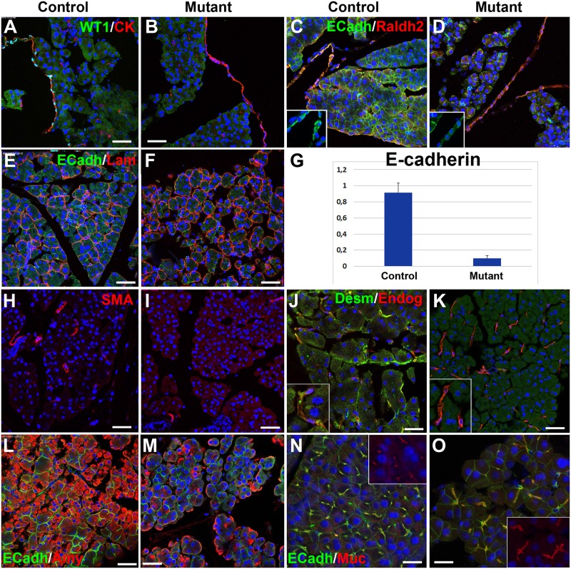
The Wilms' tumor suppressor gene (Wt1) encodes a zinc finger transcription factor that plays an essential role in the development of kidneys, gonads, spleen, adrenals and heart. Recent findings suggest that WT1 could also be playing physiological roles in adults. Systemic deletion of WT1 in mice provokes a severe deterioration of the exocrine pancreas, with mesothelial disruption, E-cadherin downregulation, disorganization of acinar architecture and accumulation of ascitic transudate. Despite this extensive damage, pancreatic stellate cells do not become activated and lose their canonical markers. We observed that pharmacological induction of pancreatitis in normal mice provokes de novo expression of WT1 in pancreatic stellate cells, concomitant with their activation. When pancreatitis was induced in mice after WT1 ablation, pancreatic stellate cells expressed WT1 and became activated, leading to a partial rescue of the acinar structure and the quiescent pancreatic stellate cell population after recovery from pancreatitis. We propose that WT1 modulates through the RALDH2/retinoic acid axis the restabilization of a part of the pancreatic stellate cell population and, indirectly, the repair of the pancreatic architecture, since quiescent pancreatic stellate cells are required for pancreas stability and repair. Thus, we suggest that WT1 plays novel and essential roles for the homeostasis of the adult pancreas and, through its upregulation in pancreatic stellate cells after a damage, for pancreatic regeneration. Due to the growing importance of the pancreatic stellate cells in physiological and pathophysiological conditions, these novel roles can be of translational relevance.


Conflict of interest statement

The authors have declared that no competing interests exist.

Figures

Fig 1
Fig 1. WT1 expression and Wt1-lineage cells in adult pancreas.
A. Wt1Cre;R26REYFP mice. Cells derived from a Wt1-expressing cell lineage WT1 show YPF expression (green). Only mesothelial cells express WT1 protein (red) (arrows). Cells derived from the Wt1-expressing cell lineage inside the pancreas do not express WT1 (arrowheads). B,C. Wt1GFP/+ reporter mice. Expression of WT1 (green cells) is restricted to the mesothelium, although some mesothelial cells do not express the reporter WT1 (arrowheads in B). Pancreatic stellate cells (PSC), characterized by desmin expression, do not express the reporter WT1 (arrowheads in C). D. RT-PCR of WT1 in mesothelium (Mes) and acinar tissue (Ac) of wildtype pancreas (+) and pancreas from mice with WT1 deletion (-). Only the mesothelium from wildtype pancreas show expression of WT1. E,F. Part of the PSC population characterized by desmin expression also shows the Wt1-lineage marker YFP in both, exocrine (white arrows) and endocrine (red arrow) pancreas. Desmin+ PSC are arranged around the polygonal acini (insert in E). Note the presence of large non-PSC, Wt1-lineage cells inside the pancreatic islets (green arrows). The white arrowhead in F shows a desmin+ perivascular cell also derived from the Wt1-expressing lineage. G. FACS analysis of disaggregated pancreatic cells enriched in PSC by centrifugation on a Nikodenz solution. PSC are identified by high levels of side scatter and violet autofluorescence due to their lipidic, retinoid-containing vesicles. Comparison of the Wt1Cre;R26REYFP mice with a control mice (YFP negative) reveals that 15.6% of the PSC express the WT1-lineage marker YFP. Scale bars: A; 25 μm; B-F: 50 μm.
Fig 2
Fig 2. Pancreatic phenotype after systemic deletion of WT1 in the Wt1CreERT2;Wt1flox model.
A,B. Control (Wt1CreERT2-;Wt1flox/+) and mutant (Wt1CreERT2+;Wt1flox/+) mice injected with tamoxifen for five days and sacrificed at the ninth day after first injection. The pancreas of the mutant mice (arrows in B) is enlarged and filled with a jelly-like matrix. C-F. Histology of control (C,E) and mutant (D,F) pancreas, H&E staining. The acinar structure of the mutant pancreas is abnormal, showing rounded shape and reduced adhesion between cells. The islets of Langerhans appear normal in the mutant (arrows in D). G-H. The mutant pancreas does not show significant increase of fibrosis as shown by trichrome (I) and Sirius red staining (J). Compare with the respective controls (G and H). Scale bars: E,F: 25 μm; C,D,G-J: 100 μm.
Fig 3
Fig 3. Pancreatic phenotype after systemic deletion of WT1 in the Wt1CreERT2;Wt1flox model.
A,B. WT1 is expressed only in the pan-cytokeratin+ mesothelial cells of control mice (A) and is deleted in the mutant mice (B). C,D. E-cadherin expression is downregulated in the pancreas of the mice with deletion of WT1, both in acini and mesothelium (inserts). However, mesothelial expression of RALDH2 does not change in mutant mice. E,F. Laminin expression is maintained in the mutant mice, but it reveals changes in the size, shape and adhesion of the acini. Note the E-cadherin immunoreactivity decrease in the mutant. G. E-cadherin downregulation in mutant mice is confirmed by qPCR (mean of three biological replicates, p<0.01, Student’s t test). H,I. Pancreatic stellate cells are not activated by the ablation of WT1, as demonstrated by the lack of SMC α-actin expression. J,K. Desmin+ pancreatic stellate cells become very scarce in the mutant mice. However, endoglin (CD105) expression is maintained in putative PSC. L,M. Amylase immunoreactivity is significantly reduced in mutant mice (quantified in Table 1). N,O. Upregulation and changes in the localization of mucin-1 immunoreactivity in mutant mice suggests loss of acinar cell polarity. Scale bars: 50 μm.
Fig 4
Fig 4. Pancreatic mesothelium is disrupted after WT1 deletion.
A. Scanning electron micrography of normal pancreatic mesothelium. B. Pancreatic mesothelium of a control mice (Wt1CreERT2;Wt1flox/+) treated with tamoxifen. C. Pancreatic mesothelium of a mutant mice (Wt1CreERT2;Wt1flox/+) showing reduced adhesion between mesothelial cells. D,E. Disruption of the mesothelium of the mutant mice (E) is shown by pan-cadherin immunolocalization. F,G. Pan-cytokeratin immunolocalization. Different morphologies are observed in the pancreatic mesothelium of these control mice, but mesothelial cells were always forming a continuous cell layer. H,I. Pan-cytokeratin immunolocalization in the mutant mice reveals disruption of the mesothelium. Scale bars: 50 μm.
Fig 5
Fig 5. Localization of cells derived from the Wt1-expressing cell lineage (Wt1Cre;R26REYFP model) after induction of pancreatitis with caerulein.
A-L show results obtained after 48 h of the induction and M-T show the pancreas after three weeks. A-D. Caerulein induced pancreatitis provokes a strong upregulation of RALDH2 and SMC α-actin in pancreatic stellate cells. Most of these activated cells belong to the Wt1-expressing cell lineage (arrowheads in B and D)), but some of them do not express the Wt1 lineage reporter (arrows in B and D). E,F. WT1 is only expressed in the mesothelium of control pancreas (arrow in E), but pancreatitis induces expression of WT1 in periacinar, putative pancreatic stellate cells (arrows in F). Note the upregulation of WT1 in the mesothelium, which is apparently releasing cells to the mesothelial space (insert). G,H. RALDH2 upregulation provoked by pancreatitis occurs in WT1+ cells, either from the Wt1 lineage (arrowheads in H) or not (arrows in H). In the latter cells WT1 is expressed de novo, as demonstrated by the lack of the lineage marker YFP. In normal mice WT1 and RALDH2 expression only coincide in the mesothelium (arrows in G). I,J. Pancreatitis induces proliferation of Wt1-lineage cells in both, mesothelium (arrow) and periacinar areas (arrowheads). K,L. Desmin expression is maintained in pancreatic stellate cells after induction of pancreatitis. Wt1 expression is upregulated in the mesothelium (arrows). The insert (only red and green channels) shows desmin+ cells expressing WT1 protein. M,N. After three weeks of pancreatitis induction, WT1 expression is again restricted to the mesothelium (arrows). Note the accumulation of submesothelial cells in the caerulein-treated mice. O,P. RALDH2 expression appears in the mesothelium (arrows) but also in submesothelial cells that are not derived from a Wt1-expressing lineage (arrowheads). Q,R. SMC α-actin, marker of activation of pancreatic stellate cells, has disappeared after three weeks, and it is not expressed by submesothelial cells (arrow). S,T. After recovery of the pancreatitis, desmin+ pancreatic stellate cells are abundant and frequently express the Wt1-lineage marker YFP (arrows in T). Scale bars: 50 μm.
Fig 6
Fig 6. Pancreatic phenotype after systemic deletion of WT1 in the Wt1CreERT2;Wt1flox model and induction of pancreatitis with caerulein.
A-F show results obtained after 48 h and G-O represent the pancreas after 10–12 days, when mice have recovered from pancreatitis. A-C. WT1 is expressed in mesothelial and stromal cells of the mouse with pancreatitis (A), it is lacking in mice after deletion of WT1 (B), but it is expressed in stromal cells after induction of pancreatitis (arrows in C). D-F. Markers of pancreatic stellate cell activation (SMC α-actin, RALDH2) are upregulated by induction of pancreatitis, even when WT1 expression has been ablated as shown in F. G-I. WT1 expression persists in mice with WT1 deletion after recovery of pancreatitis (arrows in I), and this expression appears even in some pan-cytokeratin+, mesothelial cells (arrowheads in I). J-L. The number of desmin+ expressing cells (putative stellate cells) increase in the pancreas with WT1 deletion after pancreatitis (arrows in L). These cells lose the desmin immunoreactivity in WT1-deficient mice, but they keep the CD105 expression (arrows in K) even in the islets (arrowhead). M-O. Proliferating cells are abundant in the pancreatic stroma of mice with WT1 deletion after recovery of pancreatitis (arrows in O), but they are very scarce and limited to the mesothelium in mutant mice that have not suffered of pancreatitis (arrows in N). Note the increase in RALDH2 in the mutant mice after recovery of pancreatitis (quantified in S5 Fig). Scale bars: A = 50 μm, B-O = 50 μm.

References

    1. Zha M, Li F, Xu W, Chen B, Sun Z. Isolation and characterization of islet stellate cells in rat. Islets.2014;6: e28701 10.4161/isl.28701 - DOI - PMC - PubMed
    1. Jaster R. Molecular regulation of pancreatic stellate cell function. Mol Cancer 2004;3:26 10.1186/1476-4598-3-26 - DOI - PMC - PubMed
    1. Brun PJ, Wongsiriroj N, Blaner WS. Retinoids in the pancreas. Hepatobiliary Surg Nutr. 2016;5:1–14. 10.3978/j.issn.2304-3881.2015.09.03 - DOI - PMC - PubMed
    1. Apte MV, Pirola RC, Wilson JS. Pancreatic stellate cells: a starring role in normal and diseased pancreas. Front Physiol. 2012;3:344 10.3389/fphys.2012.00344 - DOI - PMC - PubMed
    1. Haqq J, Howells LM, Garcea G, Metcalfe MS, Steward WP, Dennison AR. Pancreatic stellate cells and pancreas cancer: current perspectives and future strategies. Eur J Cancer.2014;50: 2570–2582. 10.1016/j.ejca.2014.06.021 - DOI - PubMed

Publication types

MeSH terms